Imaging Techniques To Detect Bladder Cancer
Imaging techniques, which include ultrasound, computed tomography scanning, magnetic resonance imaging and x-ray approaches, provide an important means of assessing the urinary tract, including the kidneys, and play an important role in the detection, diagnosis, and monitoring of bladder cancer.
Imaging is used in both an exploratory setting when another test suggests an anomaly, or to help confirm a diagnosis. The use of a specific imaging procedure linked to bladder cancer is dependent on a number of factors including other irregular test results, local access and availability, pre-existing medical conditions, the characteristics of a suspected tumor or unusual growth , and possible side effects of the procedure.
Irregularities in the upper urinary tract are often assessed with imaging given its accuracy in this setting and an inability to access the region via cystoscopy. In contrast, imaging techniques are less useful for diagnosing tumors in the lower urinary tract. Small and flattened bladder tumors may also be difficult to visualize with imaging.
Its important to note that imaging is generally used in combination with other bladder cancer diagnostic tests to reach a diagnosis. Cxbladder, a genomic urine test, for example, can be used with imaging to increase overall detection accuracy, and to rule out bladder cancer in low risk patients without the need for further invasive procedures.Learn more about Cxbladder
Ct Scan Imaging In Bilharziasis
The above CT images show hyperdense, extensive involvement of the urinary bladder suggesting calcification of the bladder wall. This is a typicalappearance of Schistosomiasis of the urinary bladder and is caused by calcification of the dead schistosoma parasites or their eggs. CAT scan images are courtesy of Nirmali Dutta, MD, UAE.
Producing A Sound Wave
A sound wave is typically produced by a piezoelectrictransducer encased in a plastic housing. Strong, short electrical pulses from the ultrasound machine drive the transducer at the desired frequency. The frequencies can vary between 1 and 18 MHz, though frequencies up to 50100 megahertz have been used experimentally in a technique known as biomicroscopy in special regions, such as the anterior chamber of the eye.
Older technology transducers focused their beam with physical lenses. Contemporary technology transducers use digital antenna array techniques to enable the ultrasound machine to change the direction and depth of focus. Near the transducer, the width of the ultrasound beam almost equals to the width of the transducer, after reaching a distance from the transducer , the beam width narrows to half of the transducer width, and after that the width increases , where the lateral resolution decreases. Therefore, the wider the transducer width and the higher the frequency of ultrasound, the longer the Fresnel zone, and the lateral resolution can be maintained at a greater depth from the transducer. Ultrasound waves travel in pulses. Therefore, a shorter pulse length requires higher bandwidth to constitute the ultrasound pulse.
Also Check: Pumpkin Seed Oil For Bladder Control
Urine Culture Testing To Check For Utis
Urine culture testing can be used to check for urinary tract infections.1 The symptoms of bladder cancers and urinary tract infections can be quite similar, so it is important for healthcare providers check for both infection and cancer if either could be the cause. To carry out this test, the urine sample is left in a dish in the laboratory for several days, which allows any bacteria that may be contained in the urine to grow.
Risk Factor: Chemical Exposure

Research suggests that certain jobs may increase your risk for bladder cancer. Metal workers, mechanics, and hairdressers are among those who may be exposed to cancer-causing chemicals. If you work with dyes, or in the making of rubber, textiles, leather, or paints, be sure to follow safety procedures to reduce contact with dangerous chemicals. Smoking further increases risk from chemical exposure.
Anyone can get bladder cancer, but these factors put you at greater risk:
- Gender: Men are three times more likely to get bladder cancer.
- Age: Nine out of 10 cases occur over age 55.
- Race: Whites have twice the risk of African-Americans.
Other factors at play include a family history of bladder cancer, previous cancer treatment, certain birth defects of the bladder, and chronic bladder irritation.
Don’t Miss: Bladder Infection Symptoms No Infection
Inaccurate Bladder Scan Recordings
There are several reasons why a bladder scan may be inaccurate :
- Part of the bladder extends outside of the scanned region or due to altered anatomy such as a displaced organ or prolapse
- The bladder contains substances other than water for example blood clots, mucous
- The type of conduction gel is used is wrong or inadequate
- Excessive body hair prevents conduction
- The scanner head is unclean, cracked, broken or positioned incorrectly
- The scanner head is moved while machine is in operation
- The wrong gender is identified before scanning the patient however for newer real-time scanners this is not identified as such a problem
- Poor technique is used by the health professional
- The patient is positioned incorrectly
- The patient is obese
- Ascites or fluid collection is in the abdominal cavity
- The scanners battery is flat and regular service and calibration has not been carried out as per manufacturers recommendations.
Are There Any Risks Or Side Effects
There are no known risks from the sound waves used in an ultrasound scan. Unlike some other scans, such as CT scans, ultrasound scans don’t involve exposure to radiation.
External and internal ultrasound scans don’t have any side effects and are generally painless, although you may experience some discomfort as the probe is pressed over your skin or inserted into your body.
If you’re having an internal scan and are allergic to latex, it’s important to let the sonographer or doctor carrying out the scan know this so they can use a latex-free probe cover.
Endoscopic ultrasounds can be a bit more uncomfortable and can cause temporary side effects, such as a sore throat or bloating.
There’s also a small risk of more serious complications, such as internal bleeding.
Page last reviewed: 28 July 2021 Next review due: 28 July 2024
You May Like: Ways To Strengthen Your Bladder
Bilharziasis Of The Urinary Bladder
This patient presented with lower urinary symptoms, dysuria and hematuria. Sonography of the pelvis showed thickening of the wall of the urinary bladder with extensive calcification. Theseultrasound images suggest a diagnosis of schistosomiasis or bilharziasis of the wall of the urinary bladder. Bilharziasis is a parasitic infestation which primarily involves the urinary bladder,though the liver and spleen may also be affected. The disease is caused by contact with water infested with the parasite- schistosoma and is endemic in parts of Africa . Both aboveimages are courtesy of Ravi Kadasne, MD, UAE.
What Do The Results Mean
Simple types of bladder ultrasounds, called bladder scans, can deliver immediate results. These scans are usually used only to measure the amount of urine in your bladder. A diagnostic bladder ultrasound produces more complicated images about the size, fullness, and lining of the bladder.
A doctor might understand what the ultrasound is showing, but a radiologist will typically interpret the images and write a report for your doctor to review.
The doctor will make an official diagnosis after an ultrasound based on the report from the radiologist. Apart from overactive bladder, a bladder ultrasound may also be able to help diagnose bladder cancer.
After a diagnosis, the doctor can begin treatments or therapies to help your symptoms, such as medications or pelvic floor exercises. Sometimes, more testing may be needed.
If the doctor isnt certain of your diagnosis after a bladder ultrasound, they might order other tests.
Some other tests that can be used to examine the bladder include:
- urine lab testing
You May Like: Bladder Cancer Spread To Liver
Liver/ Spleen Involvement In Schistosomiasis
his patient is a known case of Bilharziasis and ultrasound showed hepatosplenomegaly with increased echogenicity of the periportal regions of the portal veins suggesting periportal fibrosis.Fibrosis of the periportal regions of the liver is a known complication of hepatic involvement in schistosomiasis. Ultrasound images are courtesy of Ravi Kadasne, MD, UAE.
Read Also: Antibiotics Given For Bladder Infection
What Happens During An Ultrasound Scan
Most ultrasound scans last between 15 and 45 minutes. They usually take place in a hospital radiology department and are performed either by a doctor, radiographer or a sonographer.
They can also be carried out in community locations such as GP practices, and may be performed by other healthcare professionals, such as midwives or physiotherapists who have been specially trained in ultrasound.
There are different kinds of ultrasound scans, depending on which part of the body is being scanned and why.
The 3 main types are:
- external ultrasound scan the probe is moved over the skin
- internal ultrasound scan the probe is inserted into the body
- endoscopic ultrasound scan the probe is attached to a long, thin, flexible tube and passed further into the body
These techniques are described below.
Don’t Miss: Side Effects Of Bladder Cancer
What Is It And When Should It Be Used
So what is it exactly and what is it used for? It is an ultrasound machine that uses sound waves to scan a patient’s bladder to determine how much urine they have inside. This is typically done if there is suspicion that they are retaining urine which can cause serious issues such as a urinary tract infection or even bladder rupture if not treated.
Note that just because they are urinating, that doesn’t necessarily mean they aren’t retaining. Retaining simply means they aren’t completely emptying out their bladder. To see if this is the case, the patient is scanned immediately after they urinate. This is called checking their post void residual. This is one reason why it is important to keep up with their intake and output .
After a patient has had their foley catheter removed, they need to urinate within a certain amount of time. At the hospital where I am employed, that time frame is 8 hours. If they still haven’t gone by then, we encourage them to go and if they are unable to, we bladder scan them. If they are retaining, there is usually an order to reinsert of the catheter or performing a straight cath.
How much urine in the bladder is considered too much? We typically have separate parameters ordered for each individual. They usually range between 250 and 350 mL , but it really depends on the patient. Of course generally speaking, the larger the patient, the larger the bladder.
Kidney And Bladder Ultrasound

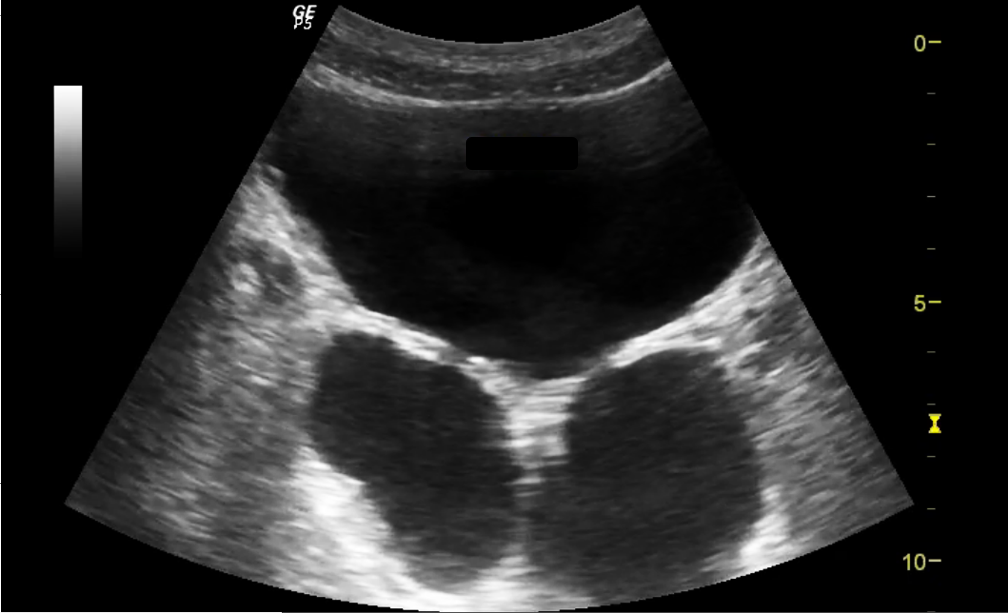
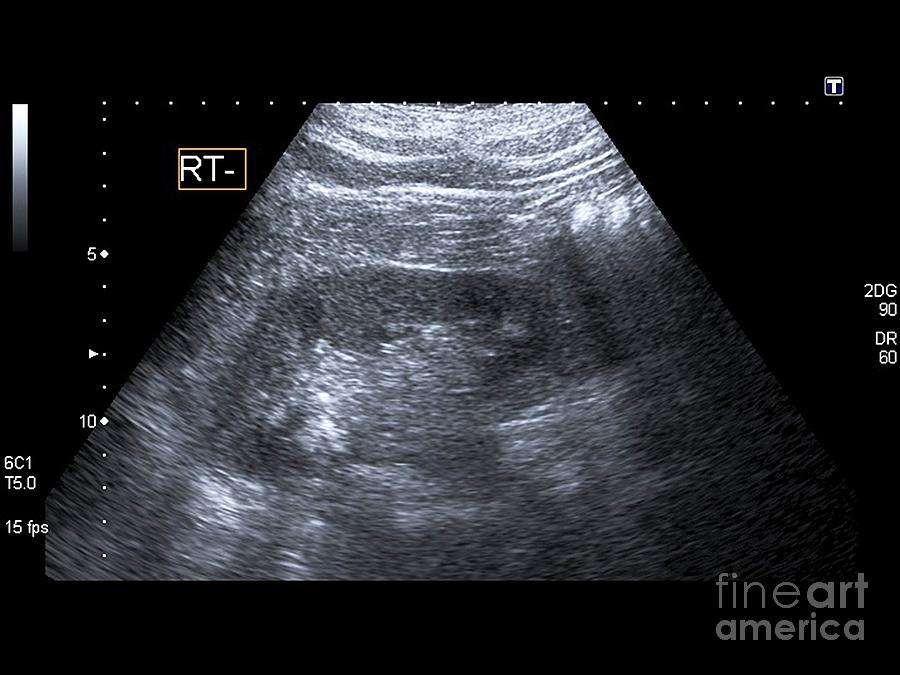
A kidney and bladder ultrasound, or renal ultrasound, uses high frequency sound waves transmitted through a transducer to visualize and assess your kidneys, ureters and urinary bladder.
Please inform the sonographer performing your exam if you have had surgery related to one of these organs, if you have a history of kidney stones or if you have stents placed in the ureters or urinary bladder.
Recommended Reading: New Drug For Overactive Bladder
Bladder Scan Results And Interpretation
The results of a bladder scan vary by patient. Bladder scan normal values form the baseline of interpreting the results of a bladder scan.
- A volume of 50ml of urine or less is considered to be adequate bladder emptying. This baseline rises to 100 ml in the elderly.
- A PVR volume of more than 200 ml is generally considered abnormal. It shows incomplete bladder emptying and warrants immediate medical attention. A PVR volume of 400 ml is considered high.
- Relief for high PVR volumes is by insertion of an in-and-out catheter.
What Happens During My Exam
- You may be asked to change into a gown.
- A warm, non-scented, hypo-allergenic ultrasound gel will be applied to the area being scanned.
- Your technologist will ask you to lower or arrange your clothing so that the area of concern is exposed to apply gel and scan the area.
- The sonographer will move the transducer around the area between your hipbones and below your belly button to take images of your kidneys and bladder.
- The sonographer will ask you to perform various breathing techniques that aid in obtaining the best images of your organs.
- You may be asked to lie on your side, sit, or stand to bring your organs into a better position.
- You may experience mild to moderate pressure while the sonographer takes the images.
- You will be asked to empty your bladder at the end of the exam and then the sonographer will take more pictures of your bladder to measure the volume of remaining urine.
- The radiologist will review the images.
- The radiologist may come into the scan room after your exam to speak to you about your results. You will then be free to leave.
You May Like: How To Fix Bladder Issues
How Do I Prepare For My Exam
- Take all prescribed medications as directed.
- Please empty your bladder 90 minutes prior to your appointment, and then drink one litre of water within the next 30 minutes. Finish drinking water one hour before your exam and do not empty your bladder.
- Please note: Drink water slowly to prevent abdominal discomfort.
- If you are too uncomfortable, please relieve your bladder of a small amount of urine.
- Once the test has begun, your sonographer will advise you if you can empty your bladder further or totally. We understand that a full bladder can be uncomfortable.
- Please arrive 15 minutes before your appointment to allow enough time to check in with reception.
- Bring photo identification and your provincial health card.
- Wear comfortable clothes.
- Please dont bring children that require supervision.
Preparing For Your Scan
Check your appointment letter for any instructions about how to prepare for your scan.
You usually need to drink about 1 litre of fluid an hour before the test, so that your bladder is comfortably full. Do not empty your bladder before the test. This is so your bladder can be seen clearly in the scan.
Take your medicines as normal unless your doctor tells you otherwise.
Also Check: How Do You Get Rid Of Overactive Bladder
What Is A Bladder Ultrasound Test
A Bladder Ultrasound Test, or Bladder Scan, is a procedure ordered by healthcare professionals when there is worry about bladder health. Specifically, the results from a bladder scan can help diagnose causes of pain or dysfunction.
In addition, the test itself is quick, safe, and painless. While bladder scans are not painful, patients will need to hold their urine so that the bladder is full for the scan.
In order to perform a bladder scan, youll need:
- A Bladder Scanner: a portable, hand-held ultrasound device that provides a virtual 3D image of the bladder quickly and the volume of urine.
- A Monitor: displays clear 3D images of the bladder from the bladder scanner.
How Bladder Scanners Work
To start, Bladder Scanners contain transducers that send out ultrasound waves. In this case, these waves bounce off the bladder to produce images before and after urination.
Then, the data is transmitted to a computerfor results. Finally, doctors and radiologists then interpret the data to diagnose and treat symptoms.
The Advantages And Precautions Of Bladder Scanning
The main benefits of bladder scanning compared with urethral catheterisation are outlined in Box 2. However, while there are many advantages, the use of a bladder scanner may not be appropriate in all situations, including if:
- The patient is morbidly obese
- There is severe abdominal scarring
- Abdominal staples or tension sutures are in situ
- There is an infected abdominal wound present.
The manufacturers recommendations should be checked for use in pregnant women and studies indicate that bladder scanning in children aged < 36 months is unreliable and should be used with caution . Bladder volumes were underestimated and Wyneski et al stated that significant volumes in neonates were undetected in some scanning machines.
Caution should also be taken for women in the early postpartum period – Lukasse et al indicated that when certain brands of scanners are compared, sometimes they give varying results.
Caution should be taken as this could hinder identification of the true anatomical detail of the bladder. In general, the use of bladder scanners does not result in any complications but one study did report potential adverse effects including skin irritation and allergic reaction to gel and padding. However, bladder scanners themselves are deemed very safe and no epidemiological studies have shown human risks .
Box 2. Benefits and advantages of bladder scanning compared with catheterisation
- No risk of urinary tract infection related to the procedure
- Non-invasive
Don’t Miss: Does Lemon Water Help Bladder Infections
Bladder Scan Procedure Protocol Results Interpretation Risks
When there is urinary retention a bladder scan is used to evaluate how much urine remains after you empty your bladder. A bladder scan procedure is comfortable and less risky than catheterization which has been used in the past.
Retention of urine in the bladder due to incomplete emptying can lead to health complications. The onset of this medically significant anomaly can be gradual or sudden. An ultrasound or CT scan is used in the bladder scan procedure.
How Do Ultrasounds Help Detect And Monitor Bladder Cancer

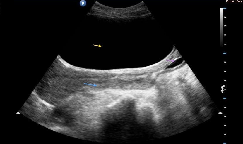
An ultrasound of the urinary tract can help assess the size of a bladder tumor and whether a bladder cancer has spread. Ultrasound is able to differentiate between fluid-filled cysts and solid tumors, however, it cannot determine if a tumor is cancerous. Ultrasound can also be used to guide a biopsy needle to sample a suspected cancer.
Ultrasound scan showing a tumor on the back wall of the bladder.
Also Check: Botox Procedure For Overactive Bladder
Bladder Ureteral Jets For Kidney Stones
Ureteral jets are a normal and periodic efflux of urine from the ureter into the bladder. Visualization of bilateral ureteral jets rules out complete obstruction of a specific ureter with high specificity .
- To see ureteral jets, scan slowly through the bladder in the transverse view and focus on the trigone .
- Turn oncolor Doppler or powerDopplerwhile scanning the bladder in the transverse view. It may take 5-10 minutes before you can visualize bilateral jets so be patient.