What Will I Experience During And After The Procedure
If your urography exam involves CT:
If the exam uses iodinated contrast material, your doctor will screen you for chronic or acute kidney disease. The doctor may administer contrast material intravenously , so you will feel a pin prick when the nurse inserts the needle into your vein. You may feel warm or flushed as the contrast is injected. You also may have a metallic taste in your mouth. This will pass. You may feel a need to urinate. However, these are only side effects of the contrast injection, and they subside quickly.
When you enter the CT scanner, you may see special light lines projected onto your body. These lines help ensure that you are in the correct position on the exam table. With modern CT scanners, you may hear slight buzzing, clicking and whirring sounds. These occur as the CT scanner’s internal parts, not usually visible to you, revolve around you during the imaging process.
You will be alone in the exam room during the CT scan, unless there are special circumstances. For example, sometimes a parent wearing a lead shield may stay in the room with their child. However, the technologist will always be able to see, hear and speak with you through a built-in intercom system.
With pediatric patients, a parent may be allowed in the room but may need to wear a lead apron to minimize radiation exposure.
If your urography exam involves MR:
Plain Films Of The Abdomen
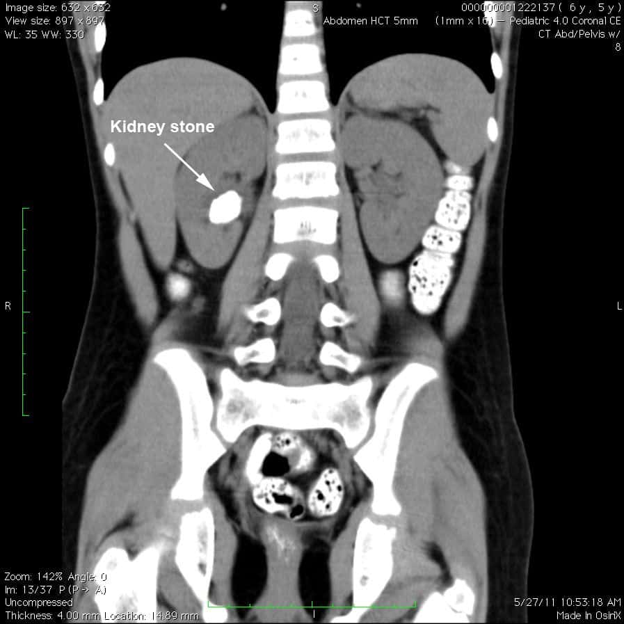
Plain films of the abdomen are now rarely used to evaluate kidney and urinary tract disease. Plain abdominal films are indicated for the evaluation of radiopaque kidney stones . An advantage of the plain film is that it can be performed in pregnant and pediatric patients, since the amount of radiation exposure is limited.
The sensitivity and specificity of plain abdominal films in detecting a stone is low in patients with renal colic and no history of kidney stones. However, plain films can be used for follow-up of stone clearance, growth, or recurrence after operative or conservative treatment of stones.
It is difficult to distinguish vascular calcifications from ureteral calcifications with plain radiography. Because of its higher sensitivity, CT imaging has replaced plain films for the diagnosis of urolithiasis and nephrolithiasis.
Plain films are not sensitive enough to exclude tumors of the kidney or urothelial tract. This imaging technique does provide general information regarding kidney size and shape.
Sound Waves Therapy: Breaking The Stones
If you are diagnosed with large stones after the CT scan for kidney stones, your doctor will recommend certain extensive treatments.
Sound waves therapy is the procedure of shock wave therapy as the strong vibrations are created to break the stones. The process usually takes fifty to sixty minutes to get completed, and you can experience some level of pain
But dont worry, your doctor might give you anesthesia so you can tolerate the treatment.
Read Also: Who To See For Bladder Problems
Ct Scan For Kidney Stones
Every year more than half a million people suffer from the issue of kidney stones. The risk of this disease has increased because of other diseases that are linked with it such as high blood pressure, diabetes, and obesity.
How do you know you have kidney stones and treatment options are available? A Ct scan for kidney stones is conducted to detect the presence of stones in your kidney.
Dive in to know more about it.
How Do Doctors Decide Which Imaging A Person Should Receive

We usually use CT first for most people, unless a tumor is much better seen on MRI. But we go back and forth as needed. If we see something on a CT scan were unsure about, we may recommend an MRI for further evaluation. If someone has several MRIs and is unable to lie still or hold their breath so we can get a good image, we may suggest a CT as an alternative. Were guided by the principle of whether the benefits of a test outweigh its risks. Thats what medical imaging is about.
You May Like: Pumpkin Seed Oil For Bladder Control
What Gets Stored In A Cookie
This site stores nothing other than an automatically generated session ID in the cookie no other information is captured.
In general, only the information that you provide, or the choices you make while visiting a web site, can be stored in a cookie. For example, the site cannot determine your email name unless you choose to type it. Allowing a website to create a cookie does not give that or any other site access to the rest of your computer, and only the site that created the cookie can read it.
How Can I Reduce My Risk For Cin And Nsf
- Know your GFR and if you have CKD. If you do not know your GFR, you can ask your doctor or healthcare professional. Your kidney function is estimated by the glomerular filtration rate, or eGFR.
- Tell all of your healthcare professionals about your GFR and CKD, especially if a diagnostic test such as a CT scan, MRI or angiogram has been ordered. Talk to the doctor ordering the diagnostic test. You can also ask to talk to the radiologist, radiology technician and nurse.
- If you need to have a CT scan, angiogram or MRI:
- Ask about your risk for NSF and CIN, based any risk factors you might have:
CIN Risk Factors
NSF Risk Factors
Use of CT scan or angiography with contrast dye, and one or more of the following:
- Heart and blood vessel problems
Use of MRI with gadolinium -based contrast dye, and one of the following:
- Advanced kidney disease
You May Like: Bladder Can T Hold Urine
Can Kidney Stones Come Back
After the kidney stone has passed or after it is removed, another stone may form. People who have had a kidney stone in the past are more likely to get another stone in the future.
If you have had a kidney stone, talk with your health care professional about your risk of getting another one. Ask your health care professional what steps you can take to lower your risk of getting another kidney stone.
What To Expect When Having A Ct Scan
A CT scan is a painless procedure that is typically performed on an outpatient basis, taking about 1030 minutes to complete.
The patient may be told not to eat or drink before the test, and a laxative or enema may be used to clear the bowels so the images are clearer. Some CT scans may be performed using special contrast dyes, which may be swallowed as a liquid, delivered intravenously, or administered with an enema. These contrast agents can help improve the quality of the CT image.
For the test, the individual lies on a flat table that slides through the middle of the scanner. Buzzing and clicking noises may be heard within the scanner. During the test, the patient may have to stay still for several minutes or to briefly hold his/her breath.
You May Like: How Long Can You Live With Untreated Bladder Cancer
What Lab Values Indicate A Need For Concern When Contrast Material Is Injected
Historically serum creatinine was the lab value used to assess kidney function. A better and more accurate measure is a lab result called estimated glomerular filtration rate . eGFR takes into account the serum creatinine value and also patient age, race and gender which affect kidney function results. At UCSF we use this very accurate blood test to assess kidney function and it can be obtained quickly, right before a scan. For CT, eGFR > 45 indicates no increased risk of kidney damage from contrast material. eGFR > 30, but less than 45 indicates that while it is safe to get contrast material, there is a small risk of causing kidney damage. In that situation, we will inject additional fluid into the patients vein before and after the contrast material injection. This hydration is effective to prevent any renal damage. For MRI, it is safe to give a regular dose of contrast material as long as the patients eGFR is > 30.
Health History And Physical Exam
Your health history is a record of your symptoms, risk factors and all the medical events and problems you have had in the past.
Your doctor will ask questions about your history of symptoms that suggest prostate cancer, such as changes in bladder habits.
Your doctor may also ask about a family history of:
- prostate cancer
- risk factors for prostate cancer
Your doctor will also do a physical exam to look for any signs of prostate cancer. During a physical exam, your doctor may:
- do a digital rectal exam to check the size and shape of the prostate and feel for any lumps or abnormal areas
- check other areas of your body, including the abdomen
You May Like: Over The Counter Bladder Medication
Recommended Reading: What Causes Bladder Pain And Pressure
What Happens After Your Imaging Test
After most imaging tests, you can go home and resume normal activity. Some tests that involve catheters may cause minor discomfort. Tests that include medication, dyes, or sedatives occasionally trigger allergic reactions.
Tests that may cause discomfort include
- Tests involving a catheter in the urethra. You might feel some mild discomfort from an irritated urethra for a few hours after the procedure.
- Transrectal ultrasound. You might feel some discomfort from an irritated rectum.
If you have a catheterization, your health care professional may prescribe an antibiotic for 1 or 2 days to prevent an infection. If you have any signs of infection, including pain, chills, or fever, call your health care professional immediately.
Tests that may cause an allergic reaction include
- Tests involving contrast medium. If you have a rare sign of reaction, such as hives, itching, nausea, vomiting, headache, or dizziness, call your health care professional immediately.
- Tests involving sedatives. If you have a rare sign of reaction, such as changes in breathing and heart rate, call your health care professional immediately.
Can An Abdomen Cat Scan Diagnose Bladder Cancer

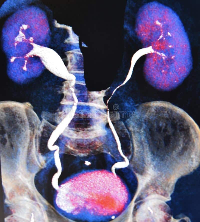
· A doctor can undertake a combined CT Scan of the kidneys, uterus, and bladder. Now, one can refer to this term as CT urogram. An individual may have detailed information about the shape and size of any tumor. Mostly, a doctor uses this to trace the tumors in the urinary tract that engulfs the bladder.
Recommended Reading: Home Remedies For Bladder Infection Pain
What Is Ultrasonography
Ultrasound uses high-frequency sound waves to create real-time images. It is a simple and painless way for urologists to look at many organs. It is flexible and offers helpful information, without using dyes or radiation.
A small probe and gel are placed on the skin. The transducer can then collect the sound waves from organs to create an image. The images look like thin, flat sections of the body on a computer screen. Newer technology can create three-dimensional ultrasound images.
These exams help your doctor diagnose many things, including organ damage. It can see the bladder, ureters and the lower part of the kidney. It is used to evaluate symptoms like:
Ultrasound is painless, safe and generally risk-free.
For more information visit our UrologyHealth.org article on ultrasounds and the different types.
What Does Imaging Mean
Imaging is a general term for techniques used to create pictures. In medicine, imaging produces pictures of bones, organs, and vessels inside the body. Imaging helps health care professionals see the cause of medical problems. Imaging techniques include
- hydronephrosis, or urine blockage, in newborns following suspicious or abnormal imaging during the pregnancy
You May Like: Dog With Bladder Cancer Passing Blood Clots
What To Expect During A Ct Urogram
During a CT urogram, you will lie on your back on a table , though you may also lie on your sides and stomach. Your healthcare provider may ask you to lie on cushions. Cushions help keep your body in the best, most comfortable position during the scan.
A healthcare provider will use a small needle and tube to deliver the contrast dye directly into a vein in your arm or hand . It usually isnt painful, but you will feel a slight pinch as the needle goes through your skin.
As the contrast dye flows through your veins, you may feel warm or flushed, almost like youre embarrassed. Some people feel nauseous or develop a headache. You may also have a salty or metallic taste in your mouth, and you may suddenly feel like you have to pee. These feelings should go away after a few moments.
When the scan begins, the bed slowly moves into the doughnut-shaped tube. Its important to stay as still as possible movement can create blurry images. Let your healthcare provider know if you have to move because youre uncomfortable or have an itch.
The scanner takes pictures of the areas that your healthcare provider needs to see. The scanner is relatively quiet as it takes pictures. Some people find the process relaxing and may fall asleep.
However, the bed may be slightly noisy as it gradually moves in and out of the scanner while taking images.
How long does a CT urogram last?
Can A Ct Scan Of The Kidneys Be Used For A Kub
Your doctor will notify you of this prior to the procedure. CT scans of the kidneys can provide more detailed information about the kidneys than standard kidney, ureter, and bladder X-rays , thus providing more information related to injuries and/or diseases of the kidneys. CT scans of the kidneys are useful in the examination
Also Check: Trouble Urinating After Bladder Sling
What Is Antegrade Pyelography
Antegrade pyelography uses a special dye to see detailed x-ray images of the upper urinary tract .
It is used to diagnose hydronephrosis, ureteropelvic junction obstruction, and obstruction of the ureters, for example.
For more information please visit our UrologyHealth.org article on Antegrade Pyelography.
Detecting Bladder Cancer With Ultrasound
An ultrasound uses high frequency sound waves to produce images of internal organs. Echoes, which are created as sound waves bounce off organs and tissues, produce computer images that provide information on the structure and movement of organs and the blood flow through vessels. An ultrasound does not use radiation or contrast dyes.
Read Also: Can Bladder Leakage Be Fixed
Tests To Find Bladder Cancer
To find bladder cancer, doctors may run tests to see whether there are certain substancessuch as bloodin the urine. Tests may include:
For patients who have symptoms or have had bladder cancer in the past, newer tests that look for tumor markers in urine may include:
- NMP22 BladderChek®
Researchers dont know yet whether these tests are reliable enough to be used for screening, but they may help find some bladder cancers.
Most doctors recommend a cystoscopy to find bladder cancer, and its often performed without anesthesia. During this procedure, the doctor inserts a long, thin tube with a camera into the urethra to see the inside of the bladder for growths and collect a tissue sample . The tissue is studied in a lab to search for cancer and obtain more information. During a cystoscopy, doctors may also perform a fluorescence cystoscopy, or blue light cystoscopy, inserting a light-activated drug into the bladder and seeing whether any cancer cells glow when they shine a blue light through the tube.
Doctors may also order imaging tests to see whether the cancer has spread. The most common imaging tests include:
Magnetic resonance imaging uses magnets and radio waves to take pictures of the inside of the body. Before the test, a contrast medium is administered orally or by injection to help make the scan clearer.
Ultrasound uses sound waves to take pictures of the inside of the body.
Read Also: Medication To Treat Bladder Infection
What Are The Reasons For A Ct Scan Of The Kidney

A CT scan of the kidney may be performed to assess the kidneys fortumors and other lesions, obstructions such askidney stones, abscesses,polycystic kidney disease, and congenital anomalies, particularly when another type ofexamination, such as X-rays or physical examination, is not conclusive.CT scans of the kidney may be used to evaluate the retroperitoneum . CT scansof the kidney may be used to assist in needle placement inkidney biopsies.
After the removal of a kidney, CT scans may be used to locate abnormalmasses in the empty space where the kidney once was. CT scans of thekidneys may be performed afterkidney transplantsto evaluate the size and location of the new kidney in relation to thebladder.
There may be other reasons for your doctor to recommend a CT scan ofthe kidney.
Also Check: How To Control My Bladder
In What Situations Is Abdominal Ct Scan Most Often Used
Abdominal and pelvic discomfort is often treated using CT scan imaging by doctors. The small intestine and colon, as well as appendicitis, pyelonephritis, and infected fluid collections, known as abscesses, may also be diagnosed using CT scan diagnostic services.
Pancreatitis, ulcerative colitis, cirrhosis of the liver, and other inflammatory bowel diseases are also detected using CT scan services.
In addition to lymphoma, abdominal CT scan detect:
- Malignancies of the ovaries and bladder.
- Urinary tract and kidney stones.
- Injury to the abdomen, such as liver, spleen, or kidneys, may develop an abdominal-abdominal aortic aneurysm.
Your doctor also prescribes abdominal CT scans to:
- Design and evaluate surgical outcomes, such as organ transplantation.
- Guides biopsies and other operations such as abscess drainages and less invasive tumor therapies.
- Staged, planned, and correctly administered Tumor radiation treatments, and the effectiveness of chemotherapy is monitored as a result.