Do Ct Pelvic Scans Detect Cancer
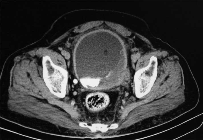
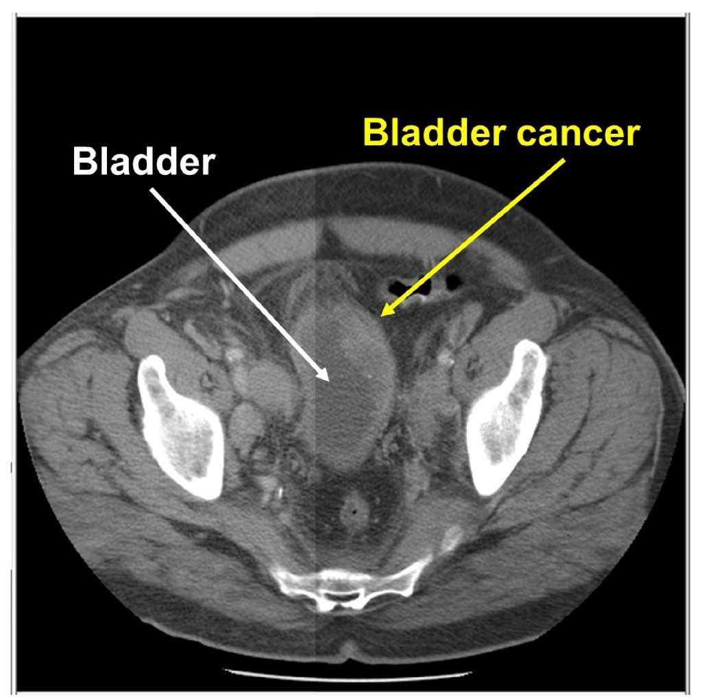
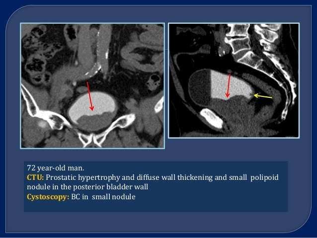
While pelvic CT scans can detect a variety of issues, they can be especially useful for detecting cancer. In particular, doctors can use this technology to look for tumors in this part of your body, but they can also use these scans to monitor the growth of tumors, to see how treatments are working, and to guide treatments.
What Will I Experience During And After The Procedure
If your urography exam involves CT:
If the exam uses iodinated contrast material, your doctor will screen you for chronic or acute kidney disease. The doctor may administer contrast material intravenously , so you will feel a pin prick when the nurse inserts the needle into your vein. You may feel warm or flushed as the contrast is injected. You also may have a metallic taste in your mouth. This will pass. You may feel a need to urinate. However, these are only side effects of the contrast injection, and they subside quickly.
When you enter the CT scanner, you may see special light lines projected onto your body. These lines help ensure that you are in the correct position on the exam table. With modern CT scanners, you may hear slight buzzing, clicking and whirring sounds. These occur as the CT scanner’s internal parts, not usually visible to you, revolve around you during the imaging process.
You will be alone in the exam room during the CT scan, unless there are special circumstances. For example, sometimes a parent wearing a lead shield may stay in the room with their child. However, the technologist will always be able to see, hear and speak with you through a built-in intercom system.
With pediatric patients, a parent may be allowed in the room but may need to wear a lead apron to minimize radiation exposure.
If your urography exam involves MR:
Transurethral Resection Of Bladder Tumor
If an abnormal area is seen during a cystoscopy, it needs to be biopsied to see if it’s cancer. A biopsy is when tiny pieces of the abnormal-looking tissue are taken out and tested for cancer cells. If bladder cancer is suspected, a biopsy is needed to be sure of the diagnosis.
The procedure used to biopsy an abnormal area is a transurethral resection of bladder tumor , also known as just a transurethral resection . During this procedure, the doctor removes the tumor and some of the bladder muscle around the tumor. The removed samples are then sent to a lab to look for cancer. If cancer is found, testing can also show if it has invaded the muscle layer of the bladder wall. For more on how this procedure is done, see Bladder Cancer Surgery.
Bladder cancer can sometimes start in more than one area of the bladder . Because of this, the doctor may take samples from many different parts of the bladder, especially if cancer is strongly suspected but no tumor can be seen. Salt water washings of the inside the bladder may also be collected and tested for cancer cells.
Read Also: How To Relieve Bladder Spasms With Catheter
Preparation For A Ct Urogram
You can usually eat and drink normally for this test. Follow the instructions given to you by your hospital.
Usually, you have to drink a certain amount of water before you arrive for your CT scan. This is so your bladder can get bigger and be seen more clearly on the pictures.
Let the radiology department know if you:
- are on medication for diabetes such as metformin
- have an allergy to contrast medium or iodine
- are pregnant or think you might be
- have any kidney problems
- are on blood thinning medicines such as warfarin
You can take all other medication as normal.
What To Expect With Bladder Ultrasounds

Ultrasounds are typically outpatient procedures and usually take 2030 minutes to complete.
Preparation is often not required ahead of an ultrasound, however, in some instances the patient will be asked to take a laxative, use an enema, or not to eat before the test. Some patients having an abdominal ultrasound may be required to drink a large amount of water so that the bladder is filled, which will improve the quality of the images.
During a bladder ultrasound, the individual is often lying down while the probe is passed over the skins surface. A lubricating gel is administered to the skin which helps the sound waves conduct.
Notably, ultrasound is a safe procedure with a low risk of side effects.
Don’t Miss: Overactive Bladder At Night Time Only
What Are The Risks Of A Ct Urogram
Radiation hazard: A single CT urogram has no risk of secondary malignancy, but multiple tests and radiation exposures may slightly increase cancer risk. However, the benefits outweigh this risk. Kidney damage: There is a small risk that the contrast medium used can affect the kidneys. The patients kidney function is checked before the test.
What Is A Cystoscopy
Its a diagnostic procedure during which a doctor inserts a cystoscope equipped with a lens into the urethra and further into the bladder.
This enables viewing of the urethra and the lining of the bladder.
Symptoms of bladder cancer have varying presentations, says Dr. Rice.
Symptoms of UTI without documented infections should be worked up by a physician to rule out anatomical abnormalities, stones and cancers.
For more information on the UTI Tracker, go to utitracker.com.
Dr. Rice is with Inova Medical Group in Fairfax, VA, and her clinical interests include bladder, kidney and prostate cancer, minimally invasive surgery and robotic surgery.
Lorra Garrick has been covering medical, fitness and cybersecurity topics for many years, having written thousands of articles for print magazines and websites, including as a ghostwriter. Shes also a former ACE-certified personal trainer.
Also Check: Causes Of Repeated Bladder Infections
How Do Ultrasounds Help Detect And Monitor Bladder Cancer
An ultrasound of the urinary tract can help assess the size of a bladder tumor and whether a bladder cancer has spread. Ultrasound is able to differentiate between fluid-filled cysts and solid tumors, however, it cannot determine if a tumor is cancerous. Ultrasound can also be used to guide a biopsy needle to sample a suspected cancer.
Ultrasound scan showing a tumor on the back wall of the bladder.
Possible Side Effects Of A Ct Scan
Sensitivity or allergic reaction to the contrast dye can occur in some patients, which may manifest as rash, nausea, wheezing, shortness of breath, or itching or swelling of the face. Symptoms are usually mild and clear on their own. Uncommon but more severe manifestations are low blood pressure and breathing difficulties.
Of additional note, while the amount of radiation used in CT scans can vary in clinical practice, CT scans deliver considerably more radiation than a typical x-ray. Therefore CT scans can carry risks associated with increased radiation exposure, such as radiation-induced future cancer.
You May Like: Losing Control Of Your Bladder
When To See A Doctor For A Ct Urogram
Your doctor may recommend a CT urogram if you are experiencing signs and symptoms, such as pain in your side or back or blood in your urine , that may be related to a urinary tract disorder. With a computerized tomography urogram, there is a small risk of an allergic reaction if the contrast material is injected.
Complete Blood Count And Chemistry Panel
On the complete blood count , the presence of anemia or an elevated white blood cell count warrants further investigation for an explanation.
The chemistry panel should include liver function studies. Although BCG is administered intravesically, systemic absorption of this agent can produce acute hepatitis. Performing baseline liver function tests before initiating therapy and repeating these tests during the course of therapy is important to help prevent serious adverse events and to determine when therapy should be stopped. In patients with suspected metastasis to liver or bone, liver function tests and measurement of the bony fraction of alkaline phosphatase should be performed.
Kidney function should be evaluated prior to the initiation of therapy because patients with marginal or abnormal kidney function may have an obstruction or some type of renal disease that may worsen with intravesical therapy. Kidney function can be evaluated with serum creatinine measurements or technetium scans of the kidneys.
Don’t Miss: What Are The Ingredients In Azo Bladder Control
How Bladder Cancer Is Diagnosed
If you or a loved one is being evaluated for bladder cancer, it can be a stressful and overwhelming time. But by learning as much as you can about the condition, including the tests performed to diagnose it, you are already taking an active role in your care.
Also, try to stay as organized as possible, be inquisitive about selecting your bladder cancer team, and attend appointments and tests with a partner or trusted loved one.
Verywell
How Is The Procedure Performed

Both CT and MR urography are usually done on an outpatient basis.
If CT urography is being performed, the technologist will begin by positioning you on the CT examination table, usually lying flat on your back or possibly on your side or stomach. You may be asked to change positions during portions of the examination. Straps and pillows may be used to help you maintain the correct position and to hold still during the exam.
Many scanners are fast enough to scan children without sedation. In special cases, children who cannot hold still may need sedation. Motion may cause blurring of the images and degrade image quality the same way that it affects photographs.
If contrast material is used, a nurse or technologist will inject the contrast through an IV line placed in the hand or arm.
Next, the table will move quickly through the scanner to determine the correct starting position for the scans. Then, the table will move slowly through the machine for the actual CT scan. Depending on the type of CT scan, the machine may make several passes.
The technologist may ask you to hold your breath during the scanning. Any motion, including breathing and body movements, can lead to artifacts on the images. This loss of image quality can resemble the blurring seen on a photograph taken of a moving object.
When the exam is complete, the technologist will ask you to wait until they verify that the images are of high enough quality for accurate interpretation by the radiologist.
Read Also: Hard To Urinate When Bladder Full
Things To Discuss With A Doctor
If a person is experiencing any urinary tract symptoms, they should contact a doctor for advice.
Without treatment, urinary tract symptoms can cause severe health issues. Likewise, untreated kidney infections can lead to problems such as kidney disease, high blood pressure, or kidney failure.
UTIs during pregnancy can also be dangerous for both the pregnant person and the fetus.
If kidney stones remain untreated, they can block the urinary tubes, which increases the risk of infection and can put a strain on the kidneys.
If a doctor recommends a CT urogram and the person has any concerns about the procedure, they should discuss the possibility of having a mild sedative. Mild sedation can help alleviate anxiety and any feelings of claustrophobia during the procedure.
Can Ct Virtual Cystoscopy Replace Conventional Cystoscopy In Early Detection Of Bladder Cancer
Ankush Jairath
1Muljibhai Patel Urological Hospital, Dr. Varendra Desai Road, Nadiad, Gujarat 387001, India
Academic Editor:
Abstract
Aim. To correlate findings of conventional cystoscopy with CT virtual cystoscopy in detecting bladder tumors and to evaluate accuracy of virtual cystoscopy in early detection of bladder cancer. Material and Method. From June 2013 to June 2014, 50 patients with history and investigations suggestive of urothelial cancer, with mean age 62.76 ± 10.45 years, underwent CTVC by a radiologist as per protocol and subsequently underwent conventional cystoscopy the same day or the next day. One urologist and one radiologist, blinded to the findings of conventional cystoscopy, independently interpreted the images, and any discrepant readings were resolved with consensus. Result. CTVC detected 23 out of 25 patients with bladder tumor correctly. Two patients were falsely detected as negative while two were falsely labeled as positive in CTVC. Virtual and conventional cystoscopy were comparable in detection of tumor growth in urinary bladder. The sensitivity, specificity, positive predictive value, and negative predictive value of virtual cystoscopy were 92% each. . CTVC correlates closely with the findings of conventional cystoscopy. Bladder should be adequately distended and devoid of urine at the time of procedure. However, more studies are required to define the role of virtual cystoscopy in routine clinical practice.
1. Introduction
2.1. Patients
Read Also: Causes Of Weak Bladder Control
How Your Urologist Would Detect Bladder Cancer
Karl Marvin Tan MD
Although urologists are commonly associated with male reproductive organisms, their specialties can be broad in scope, including the treatment of problems and disorders in urinary tracts. Your urologist also helps you detect and treat any form of bladder cancer one of the most common cancers in all.
It is said that bladder cancer is highly treated. Pelvic pain, painful urination, or blood in your urine are the main symptoms you should alert your urologist to. Especially if you are a smoker or have been older and a White Male the most damaged by bladder cancer you should be on high alert.
If you suspect that you may have bladder cancer, immediately alert your urologist to tests if you can. In myriad ways this can be done:
What To Expect When Having A Ct Scan
A CT scan is a painless procedure that is typically performed on an outpatient basis, taking about 1030 minutes to complete.
The patient may be told not to eat or drink before the test, and a laxative or enema may be used to clear the bowels so the images are clearer. Some CT scans may be performed using special contrast dyes, which may be swallowed as a liquid, delivered intravenously, or administered with an enema. These contrast agents can help improve the quality of the CT image.
For the test, the individual lies on a flat table that slides through the middle of the scanner. Buzzing and clicking noises may be heard within the scanner. During the test, the patient may have to stay still for several minutes or to briefly hold his/her breath.
Recommended Reading: Best Over The Counter Bladder Control Medication
Diagnosis Of Bladder Cancer
Diagnosis is the process of finding out the cause of a health problem. Diagnosing bladder cancer usually begins with a visit to your family doctor. Your doctor will ask you about any symptoms you have and may do a physical exam. Based on this information, your doctor may refer you to a specialist or order tests to check for bladder cancer or other health problems.
The process of diagnosis may seem long and frustrating. Its normal to worry, but try to remember that other health conditions can cause similar symptoms as bladder cancer. Its important for the healthcare team to rule out other reasons for a health problem before making a diagnosis of bladder cancer.
The following tests are usually used to rule out or diagnose bladder cancer. Many of the same tests used to diagnose cancer are used to find out how far the cancer has spread . Your doctor may also order other tests to check your general health and to help plan your treatment.
What Is A Ct Urogram With Contrast
CT urogramCTcontrastCT
What is involved in a ct urogram?
During a CT urogram, an X-ray dye is injected into a vein in your hand or arm. X-ray pictures are taken at specific times during the exam, so your doctor can clearly see your urinary tract and assess how well its working or look for any abnormalities.
Contents
Don’t Miss: Will Overactive Bladder Go Away
Transurethral Resection Of A Bladder Tumour
If abnormalities are found in your bladder during a cystoscopy, you should be offered an operation known as TURBT. This is so any abnormal areas of tissue can be removed and tested for cancer .
TURBT is carried out under general anaesthesia.
Sometimes, a sample of the muscle wall of your bladder is also taken to check whether the cancer has spread. This may be a separate operation within 6 weeks of the first biopsy.
You should also be offered a dose of chemotherapy after the operation. This may help to prevent the bladder cancer returning, if the removed cells are found to be cancerous.
See treating bladder cancer for more information about the TURBT procedure.
What Do Doctors Use Them For

Doctors use CT urograms to examine the urinary system, including the kidneys, bladder, and ureters. The ureters are the tubes that carry urine from the kidneys to the bladder.
Doctors can use CT images to see if the internal structures appear healthy and work correctly and to check for any signs of disease.
A doctor may recommend a CT urogram if a person is experiencing blood in the urine, known as hematuria, or pain in the groin or lower back.
The results of the CT urogram can help doctors diagnose conditions such as:
- kidney stones
Don’t Miss: Treatment Of Overactive Bladder In Males
Detecting Bladder Cancer With Ultrasound
An ultrasound uses high frequency sound waves to produce images of internal organs. Echoes, which are created as sound waves bounce off organs and tissues, produce computer images that provide information on the structure and movement of organs and the blood flow through vessels. An ultrasound does not use radiation or contrast dyes.
Imaging Techniques To Detect Bladder Cancer
Imaging techniques, which include ultrasound, computed tomography scanning, magnetic resonance imaging and x-ray approaches, provide an important means of assessing the urinary tract, including the kidneys, and play an important role in the detection, diagnosis, and monitoring of bladder cancer.
Imaging is used in both an exploratory setting when another test suggests an anomaly, or to help confirm a diagnosis. The use of a specific imaging procedure linked to bladder cancer is dependent on a number of factors including other irregular test results, local access and availability, pre-existing medical conditions, the characteristics of a suspected tumor or unusual growth , and possible side effects of the procedure.
Irregularities in the upper urinary tract are often assessed with imaging given its accuracy in this setting and an inability to access the region via cystoscopy. In contrast, imaging techniques are less useful for diagnosing tumors in the lower urinary tract. Small and flattened bladder tumors may also be difficult to visualize with imaging.
Its important to note that imaging is generally used in combination with other bladder cancer diagnostic tests to reach a diagnosis. Cxbladder, a genomic urine test, for example, can be used with imaging to increase overall detection accuracy, and to rule out bladder cancer in low risk patients without the need for further invasive procedures.Learn more about Cxbladder
You May Like: How Can I Relax My Bladder Naturally