What Is Muscle Invasive Bladder Cancer
Muscle invasive bladder cancer is a cancer that spreads into the detrusor muscle of the bladder. The detrusor muscle is the thick muscle deep in the bladder wall. This cancer is more likely to spread to other parts of the body.
In the U.S., bladder cancer is the third most common cancer in men. Each year, there are more than 83,000 new cases diagnosed in men and women. About 25% of bladder cancers are MIBC. Bladder cancer is more common as a person grows older. It is found most often in the age group of 75-84. Caucasians are more likely to get bladder cancer than any other ethnicity. But there are more African-Americans who do not survive the disease.
What is Cancer?
Cancer is when your body cells grow out of control. When this happens, the body cannot work the way it should. Most cancers form a lump called a tumor or a growth. Some cancers grow and spread fast. Others grow more slowly. Not all lumps are cancers. Cancerous lumps are sometimes called malignant tumors.
What is Bladder Cancer?
When cells of the bladder grow abnormally, they can become bladder cancer. A person with bladder cancer will have one or more tumors in his/her bladder.
How Does Bladder Cancer Develop and Spread?
The bladder wall has many layers, made up of different types of cells. Most bladder cancers start in the urothelium or transitional epithelium. This is the inside lining of the bladder. Transitional cell carcinoma is cancer that forms in the cells of the urothelium.
Treatment For Advanced Bladder Cancer
If bladder cancer has spread to other parts of the body, it is known as advanced or metastatic bladder cancer. You may be offered one or a combination of the following treatments to help control the cancer and ease symptoms:
- systemic chemotherapy
- radiation therapy.
Immunotherapy uses the bodys own immune system to fight cancer. BCG is a type of immunotherapy treatment that has been used for many years to treat non-muscle-invasive bladder cancer.
A new group of immunotherapy drugs called checkpoint inhibitors work by helping the immune system to recognise and attack the cancer. A checkpoint immunotherapy drug called pembrolizumab is now available in Australia for some people with urothelial cancer that has spread beyond the bladder. The drug is given directly into a vein through a drip, and the treatment may be repeated every 2 to 4 weeks for up to 2 years.
Other types of checkpoint immunotherapy drugs may become available soon.
Recommended Reading: Pumpkin Seed Extract For Overactive Bladder
What Is Cancer Staging
Staging is a way of describing where the cancer is located, if or where it has invaded or spread, and whether it is affecting other parts of the body.
Doctors use diagnostic tests to find out the cancers stage, so staging may not be complete until all of the tests are finished. Knowing the stage helps the doctor recommend the best kind of treatment, and it can help predict a patient’s prognosis, which is the chance of recovery. There are different stage descriptions for different types of cancer.
For bladder cancer, the stage is determined based on examining the sample removed during a transurethral resection of bladder tumor and finding out whether the cancer has spread to other parts of the body.
This page provides detailed information about the system used to find the stage of bladder cancer and the stage groups for bladder cancer, such as stage II or stage IV.
Don’t Miss: Where Is A Man’s Bladder Located
Cancer May Spread From Where It Began To Other Parts Of The Body
When cancer spreads to another part of the body, it is called metastasis. Cancer cells break away from where they began and travel through the lymph system or blood.
- Lymph system. The cancer gets into the lymph system, travels through the lymph vessels, and forms a tumor in another part of the body.
- Blood. The cancer gets into the blood, travels through the blood vessels, and forms a tumor in another part of the body.
The metastatic tumor is the same type of cancer as the primary tumor. For example, if bladder cancer spreads to the bone, the cancer cells in the bone are actually bladder cancer cells. The disease is metastatic bladder cancer, not bone cancer.
Bladder Cancer Stage Grouping

The results are combined to determine the stage of bladder cancer for each person. There are 5 stages: stage 0 and stages I through IV .
- Stage 0, called Papillary Carcinoma and Carcinoma in Situ, is divided into stage 0a and stage 0is, depending on the type of the tumor:
- Stage 0a : Abnormal cells are found in tissue lining the inside of the bladder. These abnormal cells, which may look like tiny mushrooms growing from the lining of the bladder, may become cancer and spread into nearby normal tissue .
- Stage 0is : A flat tumor on the tissue lining the inside of the bladder. It has not grown in toward the hollow part of the bladder, and it has not spread to the thick layer of muscle or connective tissue of the bladder .
Also Check: Ways To Treat Bladder Infection At Home
Treatment For Bladder Cancer
Treatment for bladder cancer depends on how quickly the cancer is growing. Treatment is different for non-muscle invasive bladder cancer and muscle-invasive bladder cancer.
You might feel confused or unsure about your treatment options and decisions. Its okay to ask your treatment team to explain the information to you more than once. Its often okay to take some time to think about your decisions.
When deciding on treatment for bladder cancer, you may want to discuss your options with a urologist, radiation oncologist and medical oncologist. Ask your GP for referrals.
How Do Healthcare Providers Diagnose Bladder Cancer
Healthcare providers do a series of tests to diagnose bladder cancer, including:
- Urinalysis: Providers use a variety of tests to analyze your pee. In this case, they may do urinalysis to rule out infection.
- Cytology: Providers examine cells under a microscope for signs of cancer.
- Cystoscopy: This is the primary test to identify and diagnose bladder cancer. For this test, providers use a pencil-sized lighted tube called a cystoscope to view the inside of your bladder and urethra. They may use a fluorescent dye and a special blue light that makes it easier to see cancer in your bladder. Providers may also take tissue samples while doing cystoscopies.
If urinalysis, cytology and cystoscopy results show you have bladder cancer, healthcare providers then do tests to learn more about the cancer, including:
Healthcare providers then use what they learn about the cancer to stage the disease. Staging cancer helps providers plan treatment and develop a potential prognosis or expected outcome.
Bladder cancer can be either early stage or invasive .
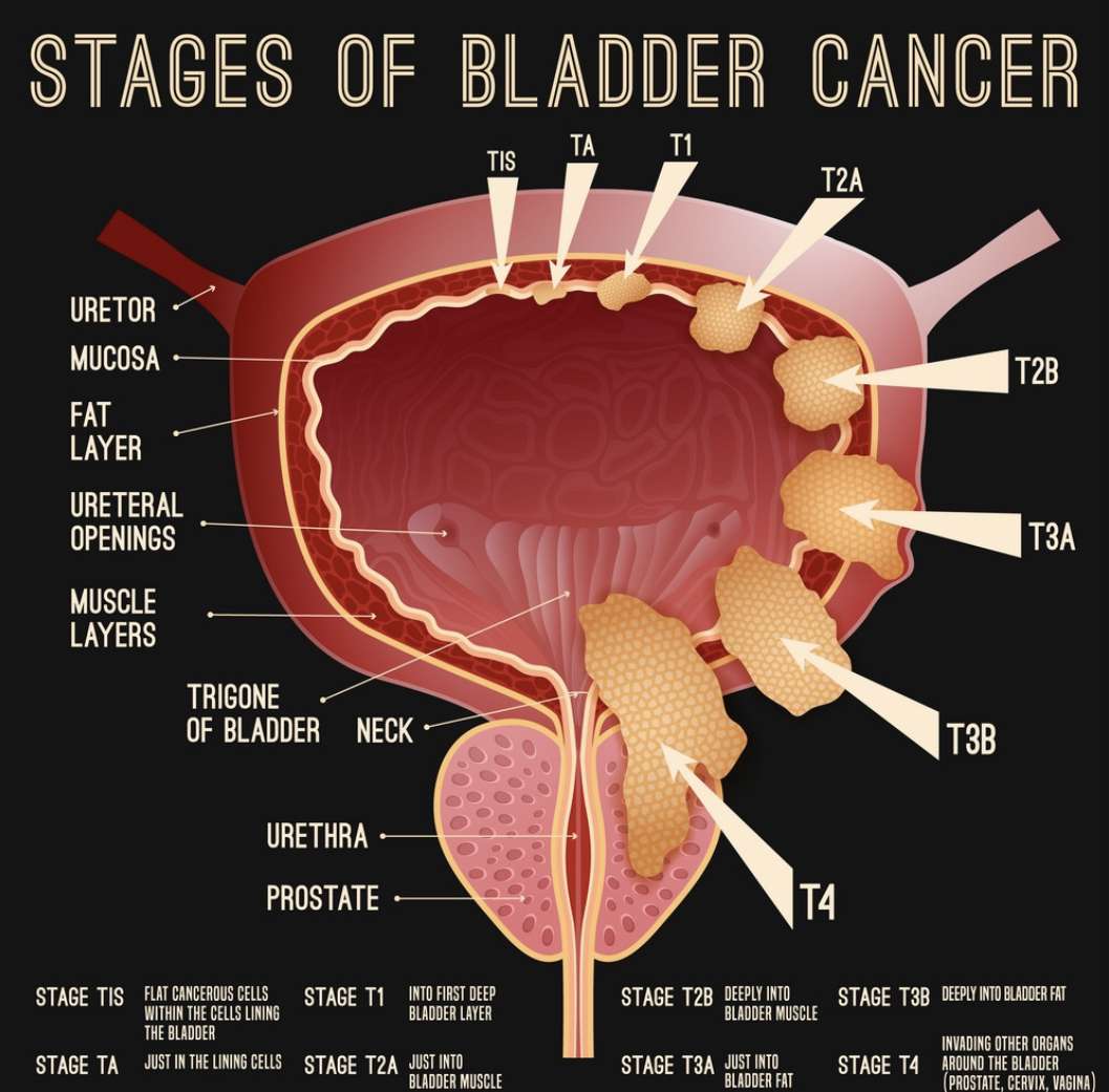
The stages range from TA to IV . In the earliest stages , the cancer is confined to the lining of your bladder or in the connective tissue just below the lining, but hasnt invaded the main muscle wall of your bladder.
Stages II to IV denote invasive cancer:
A more sophisticated and preferred staging system is TNM, which stands for tumor, node involvement and metastases. In this system:
Also Check: Neoadjuvant Chemotherapy In Bladder Cancer
Treatment Of Stages Ii And Iii Bladder Cancer
For information about the treatments listed below, see the Treatment Option Overview section.
- Transurethral resection with fulguration.
- A clinical trial of a new treatment.
Use our clinical trial search to find NCI-supported cancer clinical trials that are accepting patients. You can search for trials based on the type of cancer, the age of the patient, and where the trials are being done. General information about clinical trials is also available.
Bladder Cancer Diagnosis: Imaging
Intravenous Pyelogram
An intravenous pyelogram is an X-ray test with contrast material to show the uterus, kidneys, and bladder. When testing for bladder cancer, the dye highlights the organs of the urinary tract allowing physicians to spot potential cancer-specific abnormalities.
CT Scans and MRI
CT scans and MRI are often used to identify tumors and trace metastasized cancers as they spread to other organ systems. A CT scan provides a three-dimensional view of the bladder, the rest of the urinary tract, and the pelvis to look for masses and other abnormalities. CT scans are often used in conjunction with Positron emission tomography to highlight cells with high metabolic rates. âHot spotsâ of cells with abnormally high metabolism may indicate the presence of cancer and require further investigation.
Bone Scan
If a tumor is found in the bladder a bone scan may be performed to determine whether the cancer has spread to the bones. A bone scan involves having a small dose of a radioactive substance injected into the veins. A full body scan will show any areas where the cancer may have affected the skeletal system.
Don’t Miss: Foods To Help Bladder Infection
Non Muscle Invasive Bladder Cancer
In non muscle invasive bladder cancers, the cancer is only in the lining of the bladder. It has not grown into the deeper layers of the bladder wall. Non muscle invasive bladder cancer is also called superficial bladder cancer, or early bladder cancer.
Early bladder cancer usually appears as small growths, shaped like mushrooms. These grow out of the bladder lining. This is called papillary bladder cancer. Your surgeon can remove these growths and they may never come back.
But some types of early bladder cancer are more likely to come back. These include carcinoma in situ and high grade T1 tumours. T1 stands for the size of the tumour.
Carcinoma in situ
Unlike other early bladder cancers, areas of CIS are flat. They do not grow out of the bladder wall. In CIS the cancer cells look very abnormal and are likely to grow quickly. This is called high grade. It is more likely to come back than other types of early bladder cancer.
High grade T1 tumours
T1 tumours are early cancers that have grown from the bladder lining into a layer underneath, called the lamina propria. High grade T1 tumours are early cancers, but they can grow very quickly.
Risk groups
Doctors divide early bladder cancer into 3 risk groups. These risk groups describe how likely it is that your cancer will spread further or come back after treatment. Your risk group depends on several factors including the size of the tumour , what the tissue looks like under the microscope and type of bladder tumour.
Bladder Reconstructions And Stomas
If you have had your bladder removed, the way you pass urine will change. There are several options that your treatment team will talk to you about:
- Urostomy is where doctors create a new hole in your abdomen called a stoma. Urine drains from the stoma to the outside of your abdomen into a special bag.
- Neobladder is where a new bladder made from your small bowel forms a pouch inside your body to store urine. You will pass urine by squeezing your abdominal muscles. You will also pass a small tube into the neobladder each day to help drain the urine.
- Continent urinary diversion is a pouch made from your small bowel inside your body to store urine. The urine empties through a hole called a stoma to the outside of your abdomen into a special bag.
A bladder reconstruction is a big change in your life. You can speak with a continence or stomal therapy nurse for help, support and information. You can also call Cancer Council . You may be able to speak with a trained Cancer Council volunteer who has had cancer for tips and support.
If you find it difficult to adjust after your bladder reconstruction, it may help to be referred to a psychologist or counsellor.
Note: If you have a stoma, you can join a stoma association for support and free supplies. For more information about stoma associations, visit the Australian Council of Stoma Associations.
Also Check: Can Sugar Cause Bladder Infection
How Can I Prevent Bladder Cancer
You may not be able to prevent bladder cancer, but it may be helpful to know the risk factors that may increase the chance youll develop bladder cancer. Bladder cancer risk factors may include:
- Smoking cigarettes: Cigarette smoking more than doubles the risk of developing bladder cancer. Smoking pipes and cigars or being exposed to second-hand smoke also increases that risk.
- Cancer treatments: Radiation therapy is the second-most common risk factor. People who have certain chemotherapy drugs may also develop an increased risk of bladder cancer.
- Exposure to certain chemicals: People who work with chemicals, such as aromatic amines , are at an increased risk. Extensive exposure to rubber, leather, some textiles, paint and hairdressing supplies, typically related to occupational exposure, also appears to increase the risk.
- Infections: People who have frequent bladder infections, bladder stones or other urinary tract diseases may have an increased risk of developing bladder cancer.
- Past bladder cancer: People with a previous bladder cancer are at increased risk to form new or recurrent bladder tumors.
There Are Three Ways That Cancer Spreads In The Body

Cancer can spread through tissue, the lymph system, and the blood:
- Tissue. The cancer spreads from where it began by growing into nearby areas.
- Lymph system. The cancer spreads from where it began by getting into the lymph system. The cancer travels through the lymph vessels to other parts of the body.
- Blood. The cancer spreads from where it began by getting into the blood. The cancer travels through the blood vessels to other parts of the body.
Also Check: How To Make A Weak Bladder Stronger
Tnm Staging System For Bladder Cancer
The TNM staging system uses letters and numbers to describe the bladder cancer.
- T is how far the tumour has grown into the bladder, and how far it has spread into the surrounding tissues.
- N is whether the tumour has spread to the nearby lymph nodes.
- M is whether the tumour has spread to another part of the body .
Non-muscle-invasive bladder cancer means the cancer cells are only in the inner lining of the bladder. This means non-muscle-invasive bladder cancers are always N0 and M0.
Non-muscle-invasive bladder cancer can be staged as CIS, Ta or T1.
T Categories For Bladder Cancer
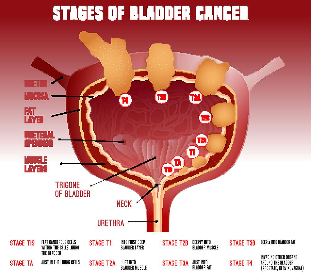
The T category describes how far the main tumor has grown into the wall of the bladder .
The wall of the bladder has 4 main layers.
- The innermost lining is called the urothelium or transitional epithelium.
- Beneath the urothelium is a thin layer of connective tissue, blood vessels, and nerves.
- Next is a thick layer of muscle.
- Outside of this muscle, a layer of fatty connective tissue separates the bladder from other nearby organs.
Nearly all bladder cancers start in the lining or urothelium. As the cancer grows into or through the other layers in the bladder, it becomes more advanced .
The T categories are described in the table above, except for:
TX: Main tumor cannot be assessed due to lack of information
T0: No evidence of a primary tumor
Read Also: Having A Hard Time Holding My Bladder
How Do I Take Care Of Myself
About half of all people with bladder cancer have early-stage cancer thats relatively easy to treat. But bladder cancer often comes back . People whove had bladder cancer will need regular checkups after treatment. Being vigilant about follow-up care is one thing you can do to take care of yourself. Here are some other suggestions from the Bladder Cancer Advocacy Network include:
- Follow a heart-healthy diet: Plan menus that include skinless poultry and fish, low-fat dairy products, nuts and legumes, and a variety of fruits and vegetables.
- Focus on high-fiber foods: Bladder cancer treatment may cause digestive issues and a fiber-rich diet may help.
- Get some exercise: Gentle exercise may help manage stress.
- Connect with others: Bladder cancer often comes back. Its not easy to have a rare disease thats likely to return. Connecting with people who understand what youre going through may help.
Urinary diversion
Some people with bladder cancer need surgery that removes their bladder and their bodies natural reservoir for pee. There are three types of urinary diversion surgeries. All three types involve surgically converting part of your intestine to become a passage tube for pee or a reservoir for storing pee.
Urinary diversion may be a challenging lifestyle change. If youll need urinary diversion surgery, ask your healthcare provider to explain each surgery types advantages and disadvantages. That way, youll know what to expect and how to take care of yourself.
Doctor Visits And Tests
Your schedule of exams and tests will depend on the stage and grade of the cancer, what treatments youve had, and other factors. Be sure to follow your doctors advice about follow-up tests.
Most experts recommend repeat exams every 3 to 6 months for people who have no signs of cancer after treatment. These are done to see if the cancer is growing back or if theres a new cancer in the bladder or urinary system. Your follow-up plan might include urine tests, physical exams, imaging tests , and blood tests. These doctor visits and tests will be done less often as time goes by and no new cancers are found.
- If your bladder hasnt been removed, regular cystoscopy exams will also be done every 3 months for at least the first 2 years.
- If you have a urinary diversion, you will be checked for signs of infection and changes in the health of your kidneys. Urine tests, blood tests, and x-rays might be used to do this. Your vitamin B12 will be checked at least once a year because urinary diversions made with your intestine can affect B12 absorption. Your doctor will also talk to you about how well youre able to control your urine. Tests will be done to look for signs of cancer in other parts of your urinary tract, too.
Some doctors recommend other lab tests as well, such as the urine tumor marker tests discussed in Can Bladder Cancer Be Found Early? Many of these tests can be used to help see if the cancer has come back, but so far none of these can take the place of cystoscopy.
Recommended Reading: What Can Cause Uncontrollable Bladder