What Is A Mr Urogram
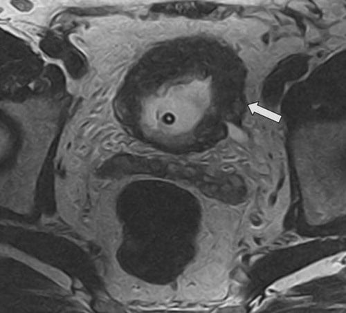
Another option for imaging is MRI of the abdomen and pelvis or MR Urogram. This test is also effective at finding tumors in the kidney and ureters and evidence of spread of cancer. It may be used to avoid radiation or in patients with contrast dye allergies or borderline kidney function. It is not quite as good at finding kidney stones and similar to CT urogram may miss tumors in the bladder such that patients still require cystoscopy.
What Are The Symptoms Of A Cancerous Cyst
Symptoms can range from mild to severe. They can include abdominal bloating and pressure, painful intercourse, and frequent urination. Some women experience menstrual irregularities, unusual hair growth, or fevers. Like noncancerous ovarian cysts, cancerous tumors sometimes cause no or only minor symptoms at first.
Ultrasound Scan For Bladder Cancer
You may have an ultrasound scan to help diagnose bladder cancer. You will need to drink plenty of fluids before the scan so your bladder is full and can be seen easier.
You may have an ultrasound scan to help diagnose bladder cancer. An ultrasound uses sound waves to create a picture of the inside of the body. It can show anything unusual in the urinary system
You will be asked to drink plenty of fluids before the test. This means your bladder is full and can be seen easier. The hospital will give you instructions about this.
When you are lying comfortably on your back, the person doing the scan spreads a gel over your tummy . A small device that makes sound waves is passed over the area. The sound waves are then turned into a picture by a computer. The scan is painless and takes about 15 to 20 minutes. Once the scan is over, you can empty your bladder.
Don’t Miss: How Do They Diagnose Bladder Cancer
Bladder Cancer Screening And Diagnosis
If doctors suspect that a patient has bladder cancer because of the presence of blood in the urine or an urgency to urinate, burning with urination, or unexplained increased frequency of urination, they may use several methods to confirm the diagnosis and determine the stage of the disease. Doctors at Columbia University Department of Urology at NewYork-Presbyterian Hospital use the latest laboratory testing and diagnostic technologies including:
Bladder Cancer Risk Factors And Warning Signs

Although the cause of bladder cancer is unknown, it is linked to tobacco use and exposure to certain chemicals.
- Smoking and Bladder Cancer: Smoking is responsible for approximately 47 percent of bladder cancer deaths among men and 37 percent among women, according to the American Cancer Society.
- Workplace Exposure and Bladder Cancer: Workers in the rubber, chemical, leather, textile, metal, and printing industries exposed to substances such as aniline dye and aromatic amines may have increased risk for bladder cancer. Other at-risk occupations include hairdressers, machinists, painters, and truck drivers.
Don’t Miss: Loss Of Bladder Control Pregnancy
Tests To Find Cancer In The Bladder
The main test to look for bladder cancer is a cystoscopy. This is an examination of the inner lining of the bladder with a cystoscope, a tube with a light and a camera on the end. Other tests can give your doctors more information about the bladder cancer. These may include an ultrasound before the cystoscopy, a biopsy taken during a cystoscopy, and a CT or MRI scan.
Learn more about:
Is Bladder Cancer Invasive Or Noninvasive
This is very important in deciding treatment. If the cancer stays in the inner layer of cells without growing into the deeper layers, its called non-invasive. If the cancer grows into the deeper layers of the bladder, its called invasive. Invasive cancers are more likely to spread and are harder to treat.
Also Check: Anticholinergic Drugs For Overactive Bladder
What Does Pelvic Ultrasound Show
In men, a pelvic ultrasound evaluates the reproductive and urinary organs . This helps the doctor find out why a man is experiencing an incomplete emptying of the urinary bladder. It can help confirm symptoms suggestive of an enlarged prostate. If you have an increased frequency of urination, your doctor may recommend a pelvic ultrasound.
Can Intestines Be Seen In Ultrasound
During the examination, an ultrasound machine sends sound waves into the abdominal area and images are recorded on a computer. The black-and-white images show the internal structures of the abdomen, such as the appendix, intestines, liver, gall bladder, pancreas, spleen, kidneys, and urinary bladder.
Don’t Miss: Why Does My Bladder Never Feel Empty
How Does Ultrasound Help With Bladder Cancer
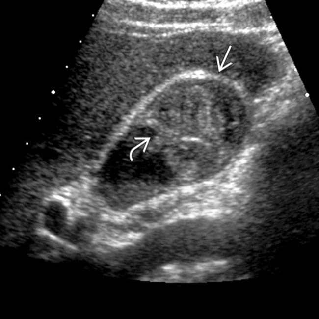
Ultrasound uses sound waves to create pictures of internal organs. It can be useful in determining the size of a bladder cancer and whether it has spread beyond the bladder to nearby organs or tissues. It can also be used to look at the kidneys. This is usually an easy test to have, and it uses no radiation.
If You Have Liver Disease
Certain diseases can make you more likely to get liver cancer, including:
- Long-term hepatitis B or C â viruses that attack and damage your liver
- Cirrhosis â liver damage that can make scar tissue replace healthy tissue
- Nonalcoholic fatty liver disease â a buildup of fat in your liver
- Liver diseases youâre born with, like hereditary hemochromatosis
Read Also: What Should I Do If I Have A Bladder Infection
Also Check: High Risk Non Muscle Invasive Bladder Cancer
How Is An Abdominal Ultrasound Done
For an abdominal ultrasound scan, you lie on your back on a comfortable table. You will need to pull up or remove your shirt or put on a hospital gown.
During the test, a trained professional:
- Applies gel to your abdomen: Water-soluble gel will cover any areas on your belly that the provider will examine. This gel may feel cold. It will not hurt you or damage your clothes.
- Moves the probe over your skin: The technician gently moves the handheld ultrasound wand over your skin, on top of the gel. The technician moves the probe back and forth until they clearly see the areas in question.
- Gives you instructions: The professional performing this test has received training in how to achieve the clearest images. They may ask you to turn to one side or hold your breath for a few seconds.
- Cleans your skin: The technician wipes off any remaining gel on your skin.
If your provider wants to study your blood vessels, your test may include Doppler ultrasound. Doppler sound waves detect details of how blood flows inside your blood vessels.
How Does Sound Work In Ultrasound

The sound waves bounce off the organs inside your body, and the microphone picks them up. The microphone links to a computer that turns the sound waves into a picture on the screen. Ultrasound scans are completely painless. You usually have the scan in the hospital x-ray department by a sonographer. A sonographer is a trained professional who is
Read Also: Essential Oils For Bladder Infection
Endoscopic Teflon Or Deflux Gel Treatment For Vesico
This patient shows an echogenic mound in the left vesico-ureteric junction. The Color Doppler image shows a ureteric jet emerging from this region suggesting that the left distal ureteric orificeis patent. This patient had a history of vesico-ureteric reflux. This was corrected by a Teflon gel injected in the submucosal part of the left VUJ viathe endoscopic route. There are 5 grades of vescio-ureteral reflux. Grade-1: the VUR reaches below the renal pelvis. Grade-2: VUR reaches up to the renal pelvis without causing dilation of thepelvis. Grade-3: There is mild to moderate dilation of the renal pelvis and ureter. Grade-4: Moderate dilation of renal pelvis, ureter and calyces is present. Grade-5: Gross dilation of pelvicalyceswith tortuous and dilated ureter. Endoscopic deflux or Teflon gel injection is used for correcting of VU reflux from grade-2 to grade-5. the gel causes a small mound to form in the submucosal part ofthe distal ureteral orifice resulting in a kind of valve formation preventing the reflux of urine up the ureter. Teflon is now being replaced by Deflux gel as the preferred material for thisprocedure. Ultrasound images of endoscopic Teflon gel injection are courtesy of Dr. ravi Kadasne, MD, UAE.
Ultrasound Images Of Urinary Bladder And Both Kidneys
Transabdominal ultrasound images show a polypoid mass in the bladder close to the bladder neck. Is this a bladder mass or an enlarged median lobe of the prostate? Faced with this dilemma wedecided to perform a transrectal ultrasound scan . The kidneys were almost normal but for a small calculus in left kidney.
Recommended Reading: Is Green Tea Good For Bladder Infection
You May Like: How Long Does Overactive Bladder Last
Can Ultrasound Detect All Stomach Problems
Doctors use ultrasound imaging tests to detect and diagnose a wide range of conditions in the human body. Many of these conditions are in the stomach and abdomen area. Ultrasounds are often used to monitor a babys health in the womb, but ultrasounds can also tell us a lot about the stomach and digestive system.
How Accurate Are Abdominal Ultrasounds
Ultrasound accuracy, as confirmed by operation, was highest for splenic masses and for aortic aneurysm . Liver masses were correctly identified in 56% of patients and gallbladder lesions in 38%. While only a 48% accuracy was obtained in diagnosing pancreatic disease, 64% of all pseudocysts were localized.
Read Also: Holistic Treatment For Overactive Bladder
What Is A Bladder Ultrasound
A bladder ultrasound is done when a doctor needs to closely examine the structure or function of your bladder.
The bladder is a muscular sac that receives urine from your kidneys, stretching to hold the fluid until you release it during urination. Bladder control, or your ability to control these muscles, makes urination a planned and purposeful task.
However, there are many issues that can complicate the process of urination.
Sediment Or Mucus In Neph Tube And Bag
I am having what looks like mucus always in my nephrostomy tube and bag. I had the tube replaced this last monday. I asked the Interventional radiologist nurse and she said it was sediment and was normal. Tonight I had some blood and talked with the resident on call and he was not sure if it was sediment or if it was mucus from my indiana pouch.
Recommended Reading: Bladder Cancer Mets To Bone
Why Did My Ultrasound Hurt
The probe will be inserted slowly and carefully, but you may still feel some discomfort as it moves. The probe will make contact with your cervix, which can feel uncomfortable for some women. You will feel some pressure as the probe is moved during the scan to take pictures from different angles.
Pelvic ultrasound and vaginal ultrasound scans can show whether:
- your ovaries are the right size
- your ovaries look normal in texture
- there are any cysts in your ovaries
Vaginal ultrasound can help to show whether any cysts on your ovaries contain cancer or not. If a cyst has any solid areas it is more likely to be cancer.
Sometimes, in women who are past their menopause, the ovaries do not show up on an ultrasound. This means that the ovaries are small and not likely to be cancerous.
If you have a suspicious looking cyst, your specialist will recommend that you have surgery to remove it. The cyst will be looked at closely in the laboratory.
Risk of malignancy index
Doctors can use a tool called the risk of malignancy index to decide if an abnormality is more likely to be cancer or not. This index combines the results of the ultrasound, CA125 blood levels and menopausal status .
This gives doctors a final score. Women with a high score are referred to a specialist multidisciplinary team . They decide on which further tests and surgery may be necessary.
Would Cancer Show Up On An Ultrasound

· A 2017 study of patients with indications for cystoscopy found that ultrasound has high sensitivity and specificity in the diagnosis of bladder cancer in patients suspected in the first stage. The most common finding was presence of papillary tumors in the bladder and the lowest frequency was related to cystic tumours.
Don’t Miss: Natural Herbal Remedies For Overactive Bladder
Tests For Bladder Cancer
Bladder cancer is often found because of signs or symptoms a person is having. Or it might be found because of lab tests a person gets for another reason. If bladder cancer is suspected, exams and tests will be needed to confirm the diagnosis. If cancer is found, more tests will be done to help find out the extent of the cancer.
Diagnosis Of Bladder Cancer
Diagnosis is the process of finding out the cause of a health problem. Diagnosing bladder cancer usually begins with a visit to your family doctor. Your doctor will ask you about any symptoms you have and may do a physical exam. Based on this information, your doctor may refer you to a specialist or order tests to check for bladder cancer or other health problems.
The process of diagnosis may seem long and frustrating. Its normal to worry, but try to remember that other health conditions can cause similar symptoms as bladder cancer. Its important for the healthcare team to rule out other reasons for a health problem before making a diagnosis of bladder cancer.
The following tests are usually used to rule out or diagnose bladder cancer. Many of the same tests used to diagnose cancer are used to find out how far the cancer has spread . Your doctor may also order other tests to check your general health and to help plan your treatment.
Recommended Reading: Why Can I Control My Bladder Female
Why Is A Bladder Ultrasound Done
About a quarter of all people in the United States experience some level of incontinence, or the inability to hold urine in the bladder until you purposely release it.
There are many causes of incontinence, and it can be difficult for a doctor to pinpoint a reason for the problem just by asking you questions or examining the outside of your body.
The following symptoms may lead a doctor to order a bladder ultrasound:
- difficulty urinating
What Is An Ultrasound
An ultrasound scan is a medical test that uses high-frequency sound waves to create live images from the inside the body. Also called a sonogram or sonography, ultrasounds let doctors see the bodys soft tissues, which X-rays cant do.
Doctors order ultrasounds for many reasons, such as to look for the causes of pain, swelling, and infection. Ultrasound scans are safe and painless.
Recommended Reading: Ways To Strengthen Your Bladder
Why Did My Abdominal Ultrasound Hurt
These waves are too high-pitched for the human ear to hear. But the waves echo as they hit a dense object, such as an organor a baby. If youre having pain in your abdomen, you may feel slight discomfort during an ultrasound. Make sure to let your technician know right away if the pain becomes severe.
Why Would A Doctor Order A Ct Scan After An Ultrasound
A CT scan may be ordered if your doctor suspects you have a tumor or blood clot. These issues could be a symptom of a very serious problem therefore the sooner they are discovered the better off the patient will be. These scans may also be used to look for signs of an infection or any excess fluid.
You May Like: Best Foods To Eat For Bladder Infection
Do I Have A Tumor In My Stomach
Feeling full: Many stomach cancer patients experience a sense of fullness in the upper abdomen after eating small meals. Heartburn: Indigestion, heartburn or symptoms similar to an ulcer may be signs of a stomach tumor. Nausea and vomiting: Some stomach cancer patients have symptoms that include nausea and vomiting.
How Do Ct Scans Help Detect And Monitor Bladder Cancer

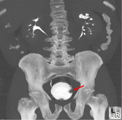
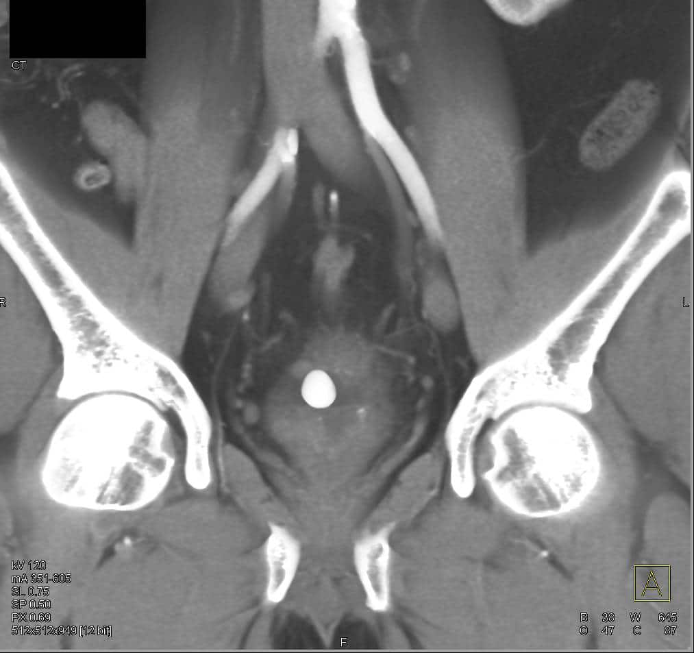
A CT urogram can:
- Determine if urinary tract abnormalities are present or if there are enlarged lymph nodes that may contain cancer
- Assess the shape, size, and location of a tumor
- Help to determine the stage of disease
- Be used to guide a biopsy needle to take samples where a bladder cancer is suspected to have spread
Computed tomography scan of the bladder showing bladder cancer .
Also Check: Why Do I Get Bladder Infection After Intercourse
Warning Signs Of Bladder Cancer
The most common warning sign of bladder cancer is blood in the urine , which may or may not be visible. Other symptoms may include: change in bladder habits, including having to urinate more often, an urgent need to urinate, or burning when you urinate needing to urinate but not being able to difficulty initiating or stopping urine flow weak, interrupted, or painful urine flow abdominal pain loss of weight or appetite persistent lower back, upper thigh, or pelvic pain.
How Much Does A Bladder Ultrasound Cost
If you have medical insurance, your copayment for a bladder ultrasound can vary or may even be free. Without insurance, the average cost of an ultrasound in the United States is between about $250 and $400.
If you have Medicare, an ultrasound may be covered under your Part A coverage if you have the procedure during an inpatient hospital stay.
At an outpatient facility, an ultrasound is covered by Medicare Part B. Your share of the cost can be between about $17 and $30 depending on where the test is done.
Recommended Reading: Colon Cancer Spread To Bladder Prognosis
What Is The Best Test For Bladder Cancer
Cystoscopy. Cystoscopy is the key diagnostic procedure for bladder cancer. It allows the doctor to see inside the body with a thin, lighted, flexible tube called a cystoscope. Flexible cystoscopy is performed in a doctors office and does not require anesthesia, which is medication that blocks the awareness of pain.