What Are Bladder Scanners
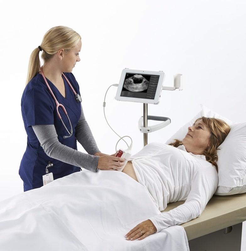
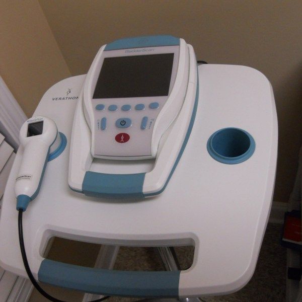
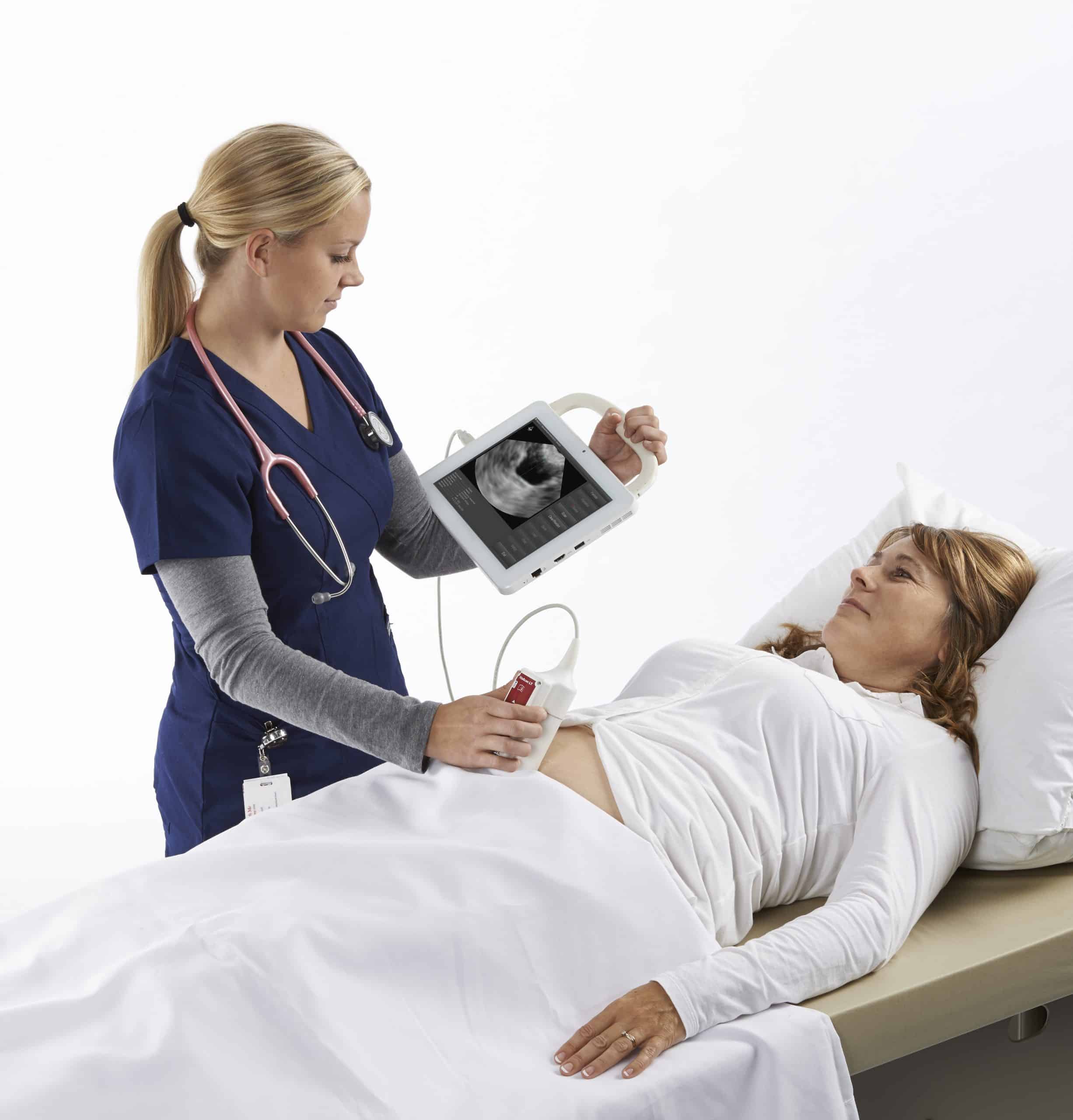
The bladder scanner is a portable, hand-held ultrasound device that can perform a quick, easy and non-invasive bladder scan. The scanner is equipped with an ultrasound probe and transducer that reflects the sound waves from the patients bladder into the scanner. The data is then transferred to a computer in a handheld unit for automatically calculating bladder volume. This ultrasonic procedure is also called Bladder Scanning or BladderScan, which is the brand name of the popular portable bladder scanner.
Bladder scanning is painless for the patient and eliminates the risks associated with unnecessary catheterization. The complete scanning lasts only a few minutes, does not require the intervention of an ultrasound technician and can prevent unnecessary invasive procedures.
What Happens During A Bladder Ultrasound
In some facilities, you may need to see a special technician for an ultrasound. But some medical offices can do this test in the examination room during a routine appointment.
Whether you have the test done in an examination room or an imaging center, the process will be similar:
Summary Of Controlled Diagnostic Experiments/ Prospective Clinical Series
Geriatric Medicine Patients and Long-Term Care Facility Patients
A community-based study by Ouslander et al. assessed the clinical utility of portable bladder ultrasound in 201 consecutive incontinent people residing in long-term care facilities who were participating in a larger clinical trial. PVR measurements were obtained using the portable bladder ultrasound model BVI 2000 or the BVI 2500 over a period of 3 years from 3 trained staff members, and then compared with the gold standard, in-and-out catheterization. The accuracy of the portable bladder ultrasound was determined by comparing the volumes obtained via catheterization and measured by bladder ultrasound.
You May Like: I Feel A Lot Of Pressure On My Bladder
How To Ultrasound The Bladder
I mentioned previously that any time a renal ultrasound is performed you should also include a bladder ultrasound. These two systems are closely linked as effect one another significantly. If you’ve spent any time in a hospital, you’ve already seen bladder ultrasound used regularly – with nursing staff performing bladder scans. This scan is simply a bladder ultrasound where the computer does all the work and calculations for you. We are going to learn today how to perform this scan manually and what information can be gathered from such a scan.
Let’s learn how!
New Technology Being Reviewed

Portable bladder ultrasound devices use automated technology to register bladder volume digitally, including PVR, and to provide three-dimensional images of the bladder. The predominant clinical use of portable bladder ultrasound is as a diagnostic aid. Health professionals administer the device to measure PVR and to prevent unnecessary catheterization. An adjunctive use of the bladder ultrasound device is to visualize the placement and removal of catheters. Also, portable bladder ultrasound products may improve the diagnosis and differentiation of urological problems and their management and treatment, including the establishment of voiding schedules, study of bladder biofeedback, reduction of UTIs, and monitoring of potential UI after surgery or trauma.
Don’t Miss: Bladder Control Exercises For Men
How Accurate Are Abdominal Ultrasounds
Ultrasound accuracy, as confirmed by operation, was highest for splenic masses and for aortic aneurysm . Liver masses were correctly identified in 56% of patients and gallbladder lesions in 38%. While only a 48% accuracy was obtained in diagnosing pancreatic disease, 64% of all pseudocysts were localized.
Read Also: Holistic Treatment For Overactive Bladder
Why Perform A Bladder Scan
The aim of the post void bladder scan is to identify if the individual is emptying their bladder effectively when voiding, or if the volume of any residual urine left is significant.
Although urinary catheterisation has long been regarded as the gold standard in determining an accurate post void residual urine volume, it is invasive, sometimes uncomfortable, and carries a risk of infection or urethral trauma. It may still be used to determine PVR if a bladder scanner is not available or a client is not suitable for bladder scanning.
Bladder scanning has advantages over many other interventions, such as intermittent catheterisation, for measurement of post void residual volume, as it is simple and causes no discomfort to the patient/client.
In order to perform bladder scanning, health professionals must undergo training and be deemed competent to perform a bladder scan.
When a bladder scanner is available, estimation of residual urine or confirmation of effective bladder emptying should be a standard component of a continence assessment.
Note: Frailty, age or dementia are not reasons to not perform a bladder scan.
Clinical Practice Point. Remember that some individuals can present with a normal voiding pattern but have a significant post void residual.
Recommended Reading: Botox Procedure For Overactive Bladder
What Is The Best Ct For Kidney Stones
CT scans of the kidneys are useful in the examination of one or both of the kidneys to detect conditions such as tumors or other lesions, obstructive conditions, such as kidney stones, congenital anomalies, polycystic kidney disease, accumulation of fluid around the kidneys, and the location of abscesses.
What are the benefits of CT scan for kidney stones?
A CT scan for kidney stones also can reduce the need for separate blood tests to aid in the diagnosis of kidney stones, so it can hasten diagnosis and treatment. The use of a spiral CT scan for kidney stones has additional benefits, including eliminating the use of contrast material to obtain images in the body.
Clinical Indicators For A Bladder Scan
For the clinician, these are:
- Part of a continence assessment
- To confirm urinary retention
- To identify incomplete bladder emptying
- To assess post void residual urine volume
- In a trial without catheter , to prevent bladder distension
- To assess bladder volume if indwelling catheter not draining
- To determine bladder volume in a client with decreased urine output.
For the patient/client, these are:
- In a bladder retraining program, it determines the need to void based on bladder volume and not time
- To assess effectiveness of mechanical bladder emptying
- To identify level of bladder sensation related to bladder volume
- To identify if adverse side effects of urinary retention are present such as with medications, e.g. anticholinergics
- As a standard investigation for at-risk groups when presenting with urinary problems
Read Also: Water Bladder Drain Pipe Cleaner
Our Purpose Is To Transform Access To Education
We offer a diverse selection of courses from leading universities and cultural institutions from around the world. These are delivered one step at a time, and are accessible on mobile, tablet and desktop, so you can fit learning around your life.
We believe learning should be an enjoyable, social experience, so our courses offer the opportunity to discuss what youre learning with others as you go, helping you make fresh discoveries and form new ideas.You can unlock new opportunities with unlimited access to hundreds of online short courses for a year by subscribing to our Unlimited package. Build your knowledge with top universities and organisations.
What Is A Bladder Scanner And What Does It Do
A Bladder Scanner can quickly and easily create a non-invasive image of the bladder. To clarify, bladder scanners have an ultrasound transducer to reflect sound waves from the patients bladder to the bladder scanner. After the transfer of data to the computer, a medical professional will analyze everything.
You May Like: Does Uti Cause Bladder Leakage
Bladder Ureteral Jets For Kidney Stones
Ureteral jets are a normal and periodic efflux of urine from the ureter into the bladder. Visualization of bilateral ureteral jets rules out complete obstruction of a specific ureter with high specificity .
- To see ureteral jets, scan slowly through the bladder in the transverse view and focus on the trigone .
- Turn oncolor Doppler or powerDopplerwhile scanning the bladder in the transverse view. It may take 5-10 minutes before you can visualize bilateral jets so be patient.
Sediment Or Mucus In Neph Tube And Bag

I am having what looks like mucus always in my nephrostomy tube and bag. I had the tube replaced this last monday. I asked the Interventional radiologist nurse and she said it was sediment and was normal. Tonight I had some blood and talked with the resident on call and he was not sure if it was sediment or if it was mucus from my indiana pouch.
Recommended Reading: Bladder Cancer Mets To Bone
Also Check: How To Live With Overactive Bladder
Tests To Find Cancer In The Bladder
The main test to look for bladder cancer is a cystoscopy. This is an examination of the inner lining of the bladder with a cystoscope, a tube with a light and a camera on the end. Other tests can give your doctors more information about the bladder cancer. These may include an ultrasound before the cystoscopy, a biopsy taken during a cystoscopy, and a CT or MRI scan.
Learn more about:
How Is An Abdominal Ultrasound Done
For an abdominal ultrasound scan, you lie on your back on a comfortable table. You will need to pull up or remove your shirt or put on a hospital gown.
During the test, a trained professional:
- Applies gel to your abdomen: Water-soluble gel will cover any areas on your belly that the provider will examine. This gel may feel cold. It will not hurt you or damage your clothes.
- Moves the probe over your skin: The technician gently moves the handheld ultrasound wand over your skin, on top of the gel. The technician moves the probe back and forth until they clearly see the areas in question.
- Gives you instructions: The professional performing this test has received training in how to achieve the clearest images. They may ask you to turn to one side or hold your breath for a few seconds.
- Cleans your skin: The technician wipes off any remaining gel on your skin.
If your provider wants to study your blood vessels, your test may include Doppler ultrasound. Doppler sound waves detect details of how blood flows inside your blood vessels.
Also Check: How Do Doctors Test For Bladder Infection
Results Of Literature Review
After scanning the literature search results, 17 articles were considered relevant to the Medical Advisory Secretariat review. Articles were found concerning the clinical utility of portable bladder ultrasound in patients who were elderly with urologic, rehabilitation, postsurgical, and neurogenic bladder conditions. All candidate articles included data on reduced or avoided urinary catheterizations plus subsequent UTI occurrences with implementation of portable bladder ultrasound. There were no RCTs found in the literature review on portable bladder ultrasound. Several of the investigation questions were not addressed by studies found in the literature review and were not included in the Medical Advisory Secretariat analysis.
What Is The Best Test For Bladder Cancer
Cystoscopy. Cystoscopy is the key diagnostic procedure for bladder cancer. It allows the doctor to see inside the body with a thin, lighted, flexible tube called a cystoscope. Flexible cystoscopy is performed in a doctors office and does not require anesthesia, which is medication that blocks the awareness of pain.
Read Also: Va Bladder Cancer Decision Granted
Measuring Bladder Volumes Scanning In The Icu
| The safety and scientific validity of this study is the responsibility of the study sponsor and investigators. Listing a study does not mean it has been evaluated by the U.S. Federal Government. Read our disclaimer for details. |
| First Posted : February 9, 2018Last Update Posted : April 3, 2019 |
| Intervention/treatment | |
|---|---|
| Urinary RetentionAcute Kidney Injury | Other: Bladder Volume Measurement bladder scanner RNOther: Bladder Volume Measurement Ultrasound APRNOther: Bladder Volume Measurement bladder scanner APRNOther: Bladder Volume Measurement Ultrasound MDOther: Intermittent Straight Catheterization |
The purpose of this correlational descriptive study is to compare measured bladder volumes with a bladder scanner , 3D ultrasound and straight catheterization in ICU patients with low urine output receiving dialysis and in ICU patients unable to void.
Upon consent of patient or LAR, patient’s age, gender and BMI with the assigned study code number will be recorded on enrollment log. Study code number, patient initials and unit will be written on bedside data collection sheet.
Sequence of 4 non-invasive measurement will vary from day to day
Data collection is complete after catheter volume is recorded.
Can Ultrasound Detect All Stomach Problems
Doctors use ultrasound imaging tests to detect and diagnose a wide range of conditions in the human body. Many of these conditions are in the stomach and abdomen area. Ultrasounds are often used to monitor a babys health in the womb, but ultrasounds can also tell us a lot about the stomach and digestive system.
Recommended Reading: Bladder Infection Keeps Coming Back
Bladder Cancer Screening And Diagnosis
If doctors suspect that a patient has bladder cancer because of the presence of blood in the urine or an urgency to urinate, burning with urination, or unexplained increased frequency of urination, they may use several methods to confirm the diagnosis and determine the stage of the disease. Doctors at Columbia University Department of Urology at NewYork-Presbyterian Hospital use the latest laboratory testing and diagnostic technologies including:
Measuring Post Void Residual Urine Volume

- A PVR of below 100ml is considered normal for any adult. This PVR may be higher in a frail older adult and cause no symptoms
- If the PVR is between 100-200ml give advice to promote more effective bladder emptying sit down with feet well supported, take time, lean forward, use double voiding. Arrange to repeat scan in 1-2 weeks.The bladder will function more effectively if the correct toilet posture and technique is followed
- If the PVR is over 200ml discuss the scan results and the individuals bladder diary information with a medical/specialist practitioner
- A PVR of 250ml is more significant if the voided volumes are low, 80-100ml than if they are higher 200-250ml because it may indicate an outflow obstruction and/or underactive bladder
- Urgent discussion is required for high post void residuals 500-1000+ ml
- If the bladder stretches to hold volumes over 1000ml the detrusor muscle is very likely to become permanently damaged, resulting in an atonic bladder.
Clinical Practice Point. Note the 1000ml is the volume voided + the residual volume. i.e. if the person voids 400ml and has a PVR of 740ml, their bladder is holding 1140ml.
Read Also: Bladder Retraining For Urge Incontinence
What Is It And When Should It Be Used
So what is it exactly and what is it used for? It is an ultrasound machine that uses sound waves to scan a patients bladder to determine how much urine they have inside. This is typically done if there is suspicion that they are retaining urine which can cause serious issues such as a urinary tract infection or even bladder rupture if not treated.
Note that just because they are urinating, that doesnt necessarily mean they arent retaining. Retaining simply means they arent completely emptying out their bladder. To see if this is the case, the patient is scanned immediately after they urinate. This is called checking their post void residual. This is one reason why it is important to keep up with their intake and output .
After a patient has had their foley catheter removed, they need to urinate within a certain amount of time. At the hospital where I am employed, that time frame is 8 hours. If they still havent gone by then, we encourage them to go and if they are unable to, we bladder scan them. If they are retaining, there is usually an order to reinsert of the catheter or performing a straight cath.
How much urine in the bladder is considered too much? We typically have separate parameters ordered for each individual. They usually range between 250 and 350 mL , but it really depends on the patient. Of course generally speaking, the larger the patient, the larger the bladder.
Recommended Reading: Can You Treat A Bladder Infection Without Antibiotics
Literature Review On Effectiveness
- Sensitivity and specificity of portable bladder ultrasound
- Effect of portable bladder ultrasound use in diagnosis between different types of UI
- Effect of portable bladder ultrasound use in diagnosis between different types of UR
- Effect of portable bladder ultrasound use in diagnosis between different types of neurogenic bladder
- Effect of portable bladder ultrasound use in diagnosis between different types of urine volume measurements, especially bladder volume and PVR urine volume
- Effect of portable bladder ultrasound use with catheterizations avoided
- Effect of portable bladder ultrasound use with discontinued catheter use
- Effect of portable bladder ultrasound use with UTIs avoided
- Effect of portable bladder ultrasound use with catheter-related damage to urinary tract
- Effect of portable bladder ultrasound use with self-toileting outcomes of bladder training
- Effect of portable bladder ultrasound use with incontinence episodes avoided
- Effect of portable bladder ultrasound use with voiding trials, voiding schedules, and biofeedback outcomes
- Effect of portable bladder ultrasound use in different settings of practice and residence
- Effect of portable bladder ultrasound use in different populations of patients by age, sex, and type of diagnosis and medical or surgical intervention
- Effect of portable bladder ultrasound use on quality of life
- Effect of portable bladder ultrasound use with nursing preference
- Harms of portable bladder ultrasound
Also Check: How To Strengthen My Bladder Muscles