Do These Blockages Always Cause Kidney Damage
No. Before birth, the mother’s placenta performs most of the functions of the kidney. As a result, babies with urinary tract abnormalities generally develop normally before delivery. In addition, many kidney or urinary tract abnormalities detected before birth have no major impact on the overall health of the baby following delivery. Nevertheless, certain conditions may interfere with the baby’s kidney function or growth after birth. For example, severely blocked flow of urine may damage the developing kidney and result in poor function after birth, a condition called dysplasia. If both kidneys are involved, the amount of urine may be seriously decreased. As a result, there may not be enough amniotic fluid surrounding the fetus, and the baby’s lungs may also be affected.
How Do I Prepare For The Test
Theres no special preparation for an ultrasound. Some pregnancy care providers ask that you come with a full bladder and dont use the restroom before the test. This helps them view your baby better on the ultrasound. You can bring a support person, but bringing children is discouraged as this is an important test that requires complete focus.
You may be asked to change into a hospital gown, but this isnt usually required for abdominal ultrasounds. If your provider is performing a transvaginal ultrasound in your first trimester, youll put on a hospital gown or undress from the waist down.
What Are Reasons You Need More Ultrasounds During Pregnancy
There are several reasons your pregnancy care provider may order additional ultrasounds during your pregnancy. Some of these reasons include:
- Problems with your ovaries, uterus, cervix or other pelvic organs.
- Your baby is measuring small for their gestational age or your provider suspects IUGR .
- Problems with the placenta like placenta previa or placental abruption.
- Youre pregnant with twins, triplets or more.
- Your baby is breech.
- You have a condition like gestational diabetes or preeclampsia.
- Your baby has a congenital disorder.
Normal results on pregnancy ultrasounds can vary. Generally, a normal result means your baby appears healthy and your provider didnt find any issues.
Don’t Miss: Does Bladder Cancer Show Up In Blood Tests
Complications Associated With Fetal Hydronephrosis
Fetal hydronephrosis is not associated with any health risks for the pregnant mother. In approximately 50% of cases, fetal hydronephrosis resolves by the third trimester. If fetal hydronephrosis gets worse as pregnancy continues, it usually does not cause any serious problems in the baby and an early delivery is not necessary. If your baby has a more severe case of fetal hydronephrosis, in-utero treatment and treatment after birth may be needed, which may include surgery to help resolve the issue and prevent long-term kidney problems.
What Do I Need To Do To Prepare For My Bladder Ultrasound

Bladder ultrasound preparation is extremely minimal. Your ultrasound technician will let you know whether you need a full or empty bladder during your test. If you need to have a full bladder your doctor may ask you to drink many glasses of water before the test. If your bladder needs to be empty your ultrasound technician will simply ask you to use the restroom before your exam. If you have trouble keeping your bladder full, you may be asked to empty your bladder about an hour before your exam and then drink water once you get to the radiologists office so that the test can be done immediately after your bladder is filled. Therefore allowing you to not have to hold your urine as long.
Don’t Miss: What Happens When A Bladder Infection Goes Untreated
What Are The Two Main Types Of Pregnancy Ultrasounds
Transvaginal ultrasound
During a transvaginal ultrasound, your pregnancy care provider places a device inside your vaginal canal . In early pregnancy, this ultrasound helps to detect a fetal heartbeat or determine how far along you are in your pregnancy . Images from a transvaginal ultrasound are clearer in early pregnancy as compared to abdominal ultrasound.
Abdominal ultrasound
Your pregnancy care provider performs an abdominal ultrasound by placing a transducer directly on your skin. Then, they move the transducer around your belly to capture images of your baby. Sometimes slight pressure has to be applied to get the best views. Providers use abdominal ultrasounds after about 12 weeks of pregnancy.
Traditional ultrasounds are 2D. More advanced technologies like 3D or 4D ultrasound can create better images. This is helpful when your provider needs to see your babys face or organs in greater detail. Not all providers have 3D or 4D ultrasound equipment or specialized training to conduct this type of ultrasound.
Your provider may recommend other types of ultrasounds. Examples of additional ultrasounds are:
Full Or Empty Bladder For Transvaginal Ultrasound
I had my first ultrasound today and the technician said it was hard to see, and said that I should empty my bladder next time but I always thought you should have a full bladder???
I dont think for transvaginal or even either for an anatomy scan, you need to have a full bladder unless your doc or the technician recommends.
they actually have me go to the bathroom prior to a transvaginal ultrasound.
Second this, even if you dont have to go they have you try lol
My tech always advised a full bladder because it extends everything in there and makes it easier to get cervical measurements and see how everything is placed, thats how she explained it to me. But for a transvaginal my tech always let me wee first because its easier to see everything that way. Your tech really should have communicated what they wanted from you before and during your procedure.
Ugh. Well next time i will make sure to go! Urg. I was measuring 6 days behind but they saw a heartbeat! I go back in a week.
They made me pee before my transvaginal, and during my anatomy scan the ultrasound tech let me get up and pee in the middle of it because she could see the fluid in my bladder. I didnt even know I had to pee but she was right she said it was for my comfort, and also my baby wasnt moving around much and we needed him to flip, and she thought maybe if I gave him some more room it would help.
Read Also: What Causes Losing Bladder Control
What Happens During A Bladder Ultrasound
In some facilities, you may need to see a special technician for an ultrasound. But some medical offices can do this test in the examination room during a routine appointment.
Whether you have the test done in an examination room or an imaging center, the process will be similar:
Diagnosis And Prognostic Criteria
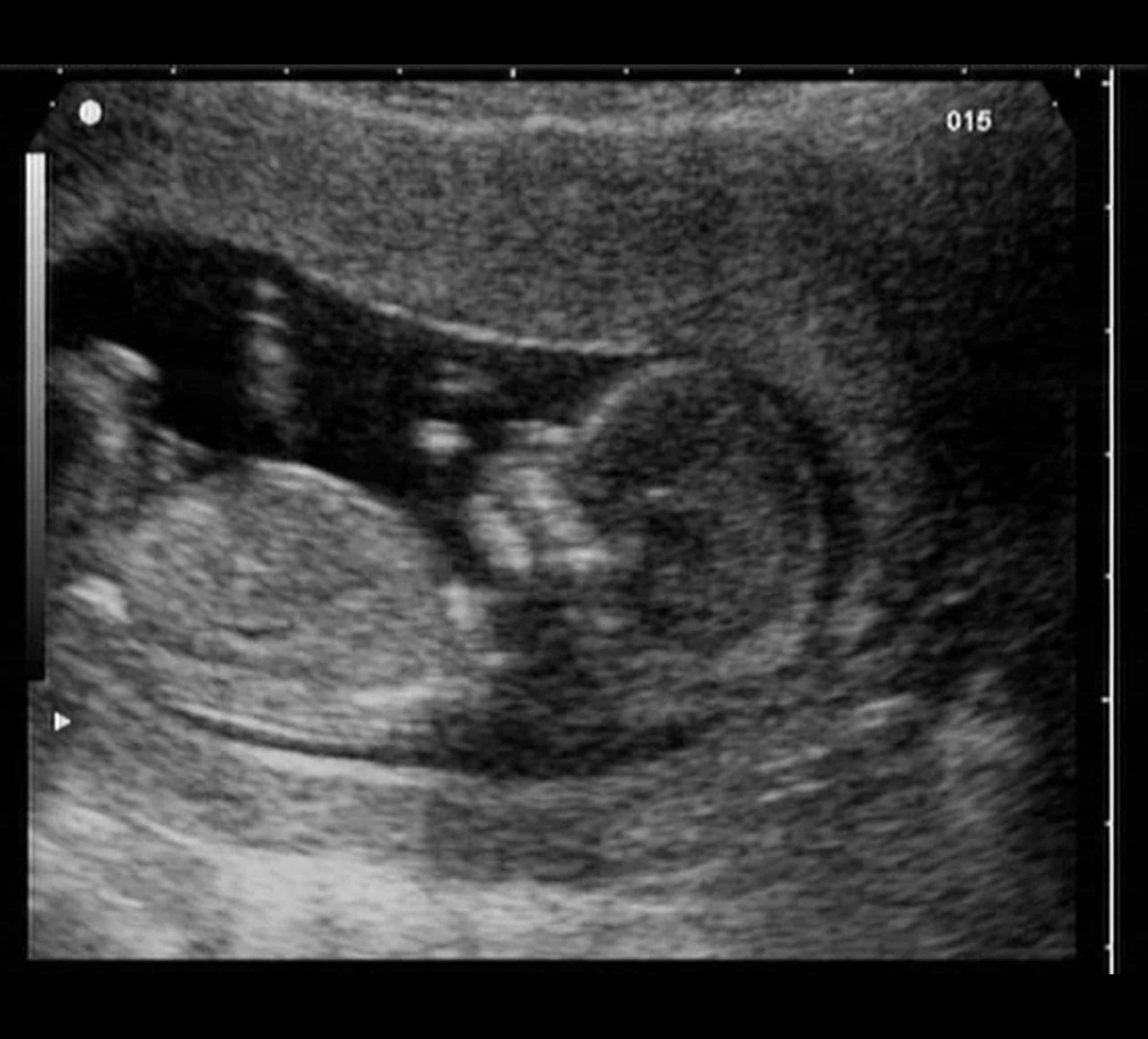
The diagnosis of LUTO is made by prenatal targeted ultrasound. Typically, the babys bladder is very distended . The presence of a key-hole sign is suggestive of posterior urethral valves, particularly in a male fetus. There may be variable degrees of dilation of the upper urinary collection system. The ultrasound findings of the babys kidneys should be carefully assessed for evidence of damage. Assessment of amniotic fluid volume as well as the presence of other potential structural abnormalities is sought. Once the diagnosis of LUTO is established, the prognosis for survival is then assessed. The babys outcomes have been correlated to the kidney function as assessed prior to treatment. There are two methods to determine the prognosis before surgery. These methods are called fetal vesicocentesis, which samples the babys urine, and cordocentesis, which samples the babys blood. Genetic studies are also performed.
You May Like: How Do Females Get A Bladder Infection
Should We Eat Before Ultrasound
You may not eat or drink anything for 8 to 10 hours before the test. If you eat, the gallbladder and ducts will empty to help digest food and will not be easily seen during the test. If your test is scheduled in the morning, we suggest that you eat nothing after midnight the night before the test is scheduled.
The Case For A Full Bladder
Fortunately, there are only a few instances of ultrasound imaging in which a full bladder is necessary:
- Renal ultrasound, or KUB ultrasound. This diagnostic test is performed to observe the kidneys and the urinary bladder. This ultrasound will also observe the seminal vesicles and prostate gland in men, and the ureters in women. The purpose of this test is to identify abnormalities in the urinary tract such as kidney stones, cysts, or tumors. Because bladder volume is measured by a renal ultrasound, it is necessary to drink plenty of water before imaging.
- Early-stage pregnancy ultrasound. When the bladder is full, the pelvic organs are easier to visualize in detail. Before week 24 of pregnancy, there is insufficient amniotic fluid to obtain the clarity needed.
In some cases, a full bladder accentuates the visibility during ultrasound imaging. In other cases, it may distort the image we need to obtain. When your doctor recommends ultrasound, the need for a full bladder may be discussed. However, if you are unsure, please contact us before your appointment. Call our Houston office at or our Humble office at We are happy to assist you in making your ultrasound as comfortable and efficient as possible.
Recommended Reading: Home Cure For Bladder Infection
British Columbia Specific Information
Your health care provider may request that you have one or more fetal ultrasound scans during your pregnancy. A fetal ultrasound scan is a medical procedure which uses sound waves to produce images of your baby in the womb. These images are used to determine the health and well-being of your baby. To learn more, see HealthLinkBC File #116 Fetal Ultrasound.
But Why A Full Bladder

Pelvic Ultrasound Full Bladder
Blame the laws of Ultrasound Physics for this one. Sound waves travel more easily through fluid than tissue. Think of your pelvic anatomy from front to back. First is skin, then fat, then muscle, then intestines or bowel, then your bladder. Your uterus sits behind your bladder. So, if you drink lots of water and fully distend your bladder, it provides a window to the uterus. Also, your bowel contains air and gas which can limit what we see. Filling your bladder pushes the intestines aside. Its actually kinda cool, but not so much if youre the one doing the drinking.
Additionally, the uterus of most women tilts forward or toward the front of your belly. Filling the bladder aids in pushing the uterus backward a littlenot up or higher, as Ive read in some pregnancy books or sites, though that phrasing is really just a technicality. When the top of the uterus tilts back a little, a better angle is created to see more clearly. Occasionally, a uterus decides to go rogue and tilts too far backward instead . Sometimes it tilts so far back that it folds over on itself . This is a totally normal variant. Often, however, the full bladder only helps minimally in these circumstances.
Don’t Miss: Can Bladder Infection Cause Kidney Infection
What Results Do You Get On A Pregnancy Ultrasound
Your ultrasound results will be normal or abnormal. A normal result means your pregnancy care provider didnt find any problems and that your baby is growing and developing normally. An abnormal result means your provider noticed something irregular. If they do, your provider will order additional ultrasounds or diagnostic tests to determine if something is wrong.
Occasionally, the ultrasound is incomplete if theres difficulty seeing all the structures needed for that particular ultrasound. Your babys position or movement sometimes makes it difficult to see everything your provider needs to see. If this is the case, youll need a repeat ultrasound and theyll try again.
There are some limitations to ultrasounds, so your provider may not find certain abnormalities until after birth.
What Causes Urinary Tract Dilation
As with most birth defects, the cause of urinary tract dilation is unknown. The condition appears to run in some families, so itâs not unusual for a parent, sibling, or cousin to have had urinary tract dilation or some other kidney issue in childhood. In rare instances, urinary track dilation is due to a genetic or chromosomal condition, such as Down syndrome . Most cases of urinary tract dilation develop in otherwise healthy babies, however.
You May Like: How To Train A Weak Bladder
Renal Pelvis Dilation And Other Indicators Predictive Of Congenital Anomalies
The timing of prenatal ultrasonography is critical to detection of congenital anomalies of the kidney and the urinary tract. The fetal kidney can be visualized on ultrasound at 12 to 15 weeks gestation, but significant disease may be difficult to detect before differentiation of the renal cortex and the medulla, which occurs at 20 to 25 weeks gestation. Ultrasound allows measurement of the anterior-posterior renal pelvis diameter and evaluation of calyceal dilation, renal parenchymal thickness and appearance, and abnormalities of the bladder or ureters. Although nephrogenesis is complete by 36 weeks gestation, the impact of continuing compression of the renal parenchyma on the ultimate endowment of healthy nephrons is unknown.
Anterior-posterior RPD measured in the transverse plane provides an index of hydronephrosis severity. An abnormal RPD has been defined in the National Institute of Child Health and Human Developments 2014 Executive Summary on Fetal Imaging as 4 mm or greater in the second trimester and 7 mm or greater at 32 weeks or more, with postnatal radiographic evaluation recommended.
Will My Baby Need An Operation
In many cases, no treatment is needed, as the condition resolves on its own without any long-lasting medical problems. An antibiotic is sometimes prescribed, however, to prevent a urinary tract infection.
If your baby has advanced urinary tract dilation , surgery may be needed to correct the underlying structural problem that is blocking the normal flow of urine. To prevent lasting kidney damage, the surgery is usually done before the childâs second birthday.
You May Like: How To Test For Bladder Infection
What Is A Pelvic Ultrasound
Ultrasound uses a transducer that sends out ultrasound waves at a frequency too high to be heard. The ultrasound transducer is placed on the skin, and the ultrasound waves move through the body to the organs and structures within. The sound waves bounce off the organs like an echo and return to the transducer. The transducer processes the reflected waves, which are then converted by a computer into an image of the organs or tissues being examined.
The sound waves travel at different speeds depending on the type of tissue encountered fastest through bone tissue and slowest through air. The speed at which the sound waves are returned to the transducer, as well as how much of the sound wave returns, is translated by the transducer as different types of tissue.
An ultrasound gel is placed on the transducer and the skin to allow for smooth movement of the transducer over the skin and to eliminate air between the skin and the transducer for the best sound conduction.
Another type of ultrasound is Doppler ultrasound, sometimes called a duplex study, used to show the speed and direction of blood flow in certain pelvic organs. Unlike a standard ultrasound, some sound waves during the Doppler exam are audible.
Pelvic ultrasound may be performed using one or both of 2 methods:
The type of ultrasound procedure performed depends on the reason for the ultrasound. Only one method may be used, or both methods may be needed to provide the information needed for diagnosis or treatment.
Baby Has Full Bladder 35 Weeks
I am currently 35 weeks two days I went for my routine appointment today. The baby has a very full bladder one that the doctor hasn’t seen in years he said. I’m a first time mom so I’m obviously very concerned but he seemed to brush it off. I’m personally not happy with this doctor and had tried switching in the second trimester but no one would take me as I was past the marker of transfer. Has anyone else ever had the issue of their baby having a very full bladder? I asked him how will we know if she’s okay and he said she would be checked upon being born and he said if necessary a valve would be opened which I’m unsure what that means. I’m really just worried for my little one like I said I’m a first time mom and this doctor I just feel always gives me the run around so it makes me even more nervous. I never had an Anatomy scan because he never gave me one and also I had the quad screening done in the second trimester which gave my results of Down syndrome as 1 in 93 so idk if this could play a role also as I didn’t have the amino done.
Don’t Miss: Does D Mannose Help Overactive Bladder