Is A Ct Scan And Ct Scan The Same
So, CAT and CT scans both mean the same type of diagnostic examination. CAT was used earlier in its history, while CT is the recent up-to-date term for convenience sake. The term CT stands for computed tomography and the term CAT stands for computed axial tomography or computerized axial tomography scan.
How Ezra Uses Mris
At Ezra, we leverage the advantages of MRI scans using state-of-the-art equipment and the expertise and experience of our dedicated healthcare team to make cancer screening fast, accurate, and affordable.
Our MRI protocol is in its sixth generation, has been validated through over 400 test scans that were verified for standard of cancer from 5 radiologists. We use 3T strength magnets and multiparametric MRI for our imaging to increase specificity and minimize false positives of cancer findings.
The Ezra Scan.
The Ezra Scan is a full-body scan service designed to screen for cancer in up to 14 internal organs. Our full body imaging provides a holistic approach to screening for early cancer as were able to look at findings across the body to detect abnormalities. It is a 60-minute MRI-based scan and a 5-minute low-dose CT of the chest that includes an optional follow-up consultation with a Medical Provider to walk through your results.
So far, 72% of our members have had clinically significant findings from our scan1. Throughout your entire Ezra experience, youll be guided by a personal Care Advisor. Your Ezra Report includes recommendations on your findings, including lifestyle and dietary changes as well as follow up steps for any signs of possible cancer.
The Ezra MRI Protocol.
The Ezra Full Body.
At $1,950 or $180 a month, the Ezra Full Body screens for potential cancers in the head, neck, abdomen, and pelvis. Here is a list of the organs covered from our Full Body:
Early Detection And Screening
Because bladder cancer causes urinary symptoms such as blood in the urine, it may be found early. However, because blood in urine is caused by a lot of conditions other than cancer, urinalysis isnt a useful screening test for the general population.
There isnt a test yet that is able to screen the general population for bladder cancer. Doctors may recommend specific tests to screen for bladder cancer based on known risk factors.
Expert
Don’t Miss: Is Heat Good For Bladder Infection
What Is A Bladder Ct Scan
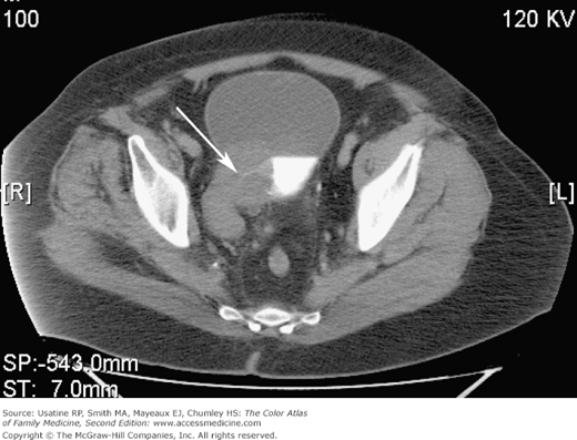
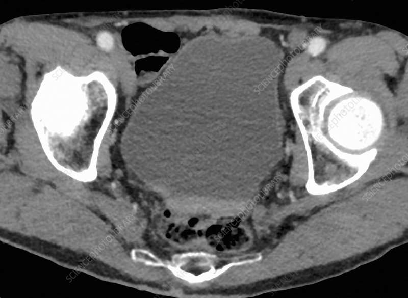
A bladder Computed Tomography scan provides an image of the bladder to help medical professionals learn more about a mass, blockage, or other problem. It involves a series of individual X-ray images, called slices, that are pulled together by a computer to provide a detailed image of the inside of the body. This test may be ordered for diagnostic reasons to determine why a patient is experiencing urinary problems, or as a follow-up to see how well a patient is responding to treatment. Imaging of other parts of the urinary tract, like the ureters and kidneys, may take place at the same time as a bladder CT scan.
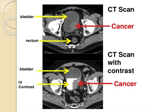
This test may be performed with the assistance of a contrast agent to highlight certain structures in the urinary tract, which is injected before the test. The patient may need a series of images taken before contrast, followed by another set after. A bladder CT scan involves lying on a table or platform inside the scanning equipment and holding still while the images are acquired. Patients concerned about claustrophobia may receive sedatives to stay relaxed and calm during the test.
Tests For Bladder Cancer

Bladder cancer is often found because of signs or symptoms a person is having. Or it might be found because of lab tests a person gets for another reason. If bladder cancer is suspected, exams and tests will be needed to confirm the diagnosis. If cancer is found, more tests will be done to help find out the extent of the cancer.
Recommended Reading: Can Bladder Infection Heal On Its Own
Transurethral Resection Of Bladder Tumor
If an abnormal area is seen during a cystoscopy, it needs to be biopsied to see if it’s cancer. A biopsy is when tiny pieces of the abnormal-looking tissue are taken out and tested for cancer cells. If bladder cancer is suspected, a biopsy is needed to be sure of the diagnosis.
The procedure used to biopsy an abnormal area is a transurethral resection of bladder tumor , also known as just a transurethral resection . During this procedure, the doctor removes the tumor and some of the bladder muscle around the tumor. The removed samples are then sent to a lab to look for cancer. If cancer is found, testing can also show if it has invaded the muscle layer of the bladder wall. For more on how this procedure is done, see Bladder Cancer Surgery.
Bladder cancer can sometimes start in more than one area of the bladder . Because of this, the doctor may take samples from many different parts of the bladder, especially if cancer is strongly suspected but no tumor can be seen. Salt water washings of the inside the bladder may also be collected and tested for cancer cells.
What Are Some Disadvantages Of Each Imaging Method
Because CTs use ionizing radiation, they could damage DNA and may very slightly raise the risk of developing cancer. The Food & Drug Administration estimates that the extra risk of any one person developing a fatal cancer from a typical CT procedure is about 1 in 2,000. MRIs do not use ionizing radiation, so there is no issue of raising cancer risk. But they take much longer to complete than CTs. MRIs require the person to lie still within a closed space for about 20 to 40 minutes. This can affect some people with claustrophobia, and the procedure is noisy, which is why we provide ear protection.
Both CT and MRI commonly require the injection of a contrast dye before or during the procedure. This helps the radiologist see organs and other tissues within the body more clearly.
Also Check: How Do Doctors Test For Bladder Infection
Use In Men Already Diagnosed With Prostate Cancer
The PSA test can also be useful if you have already been diagnosed with prostate cancer.
- In men just diagnosed with prostate cancer, the PSA level can be used together with physical exam results and tumor grade to help decide if other tests are needed.
- The PSA level is used to help determine the stage of your cancer. This can affect your treatment options, since some treatments are not likely to be helpful if the cancer has spread to other parts of the body.
- PSA tests are often an important part of determining how well treatment is working, as well as in watching for a possible recurrence of the cancer after treatment .
Flexible Cystoscopy And Biopsy
During a flexible cystoscopy, the doctor applies a local anesthetic and then inserts a cystoscope, a small flexible tube containing a tiny, lighted video camera on the end, into the urethra. He or she guides the cystoscope into the bladder to view the organs inner lining.
If the doctor sees unusual tissue or growths, a biopsy may be performed. During a biopsy, tissue is removed for examination under a microscope, so that the doctor can determine whether cancer is present. Cystoscopy can cause minor irritation of the bladder, which may result in some bleeding shortly afterward.
NYU Langone is a leader in using blue light cystoscopy, an advanced imaging procedure that helps to better detect and treat noninvasive bladder cancer tumors. Our surgeons perform this procedure using a light-sensitive contrast solution that is injected into the bladder through a catheter, or hollow tube, placed in the urethra.
After this solution accumulates in bladder cancer cells, our doctors place a cystoscope with a special blue light on the end into the bladder. The contrast solution makes the cancer cells glow brightly under the blue lightenabled cystoscope. This allows surgeons to ensure more complete tumor removal, reducing the chances of a recurrence or the need for repeat surgeries.
Recommended Reading: Does A Bladder Infection Cause Incontinence
Radiomics Features From Paired Rois
Gray-level and texture features were extracted from the paired ROIs used for the DL-CNN. Thirty-eight features, including gray-level histogram statistics, and run length statistics features, were calculated for every ROI. The per-lesion features were generated by averaging the feature values among the ROIs associated with the lesion. Similar to the RF-SL model, feature selection was performed and a random forest classifier using 2 trees and the minimum number of observations per tree leaf set of 29 was trained to use the selected radiomics features to predict the likelihood of the post-treatment lesion being T0 stage. The parameters were selected experimentally using the training set.
Health History And Physical Exam
Your health history is a record of your symptoms, risk factors and all the medical events and problems you have had in the past.
Your doctor will ask questions about your history of symptoms that suggest prostate cancer, such as changes in bladder habits.
Your doctor may also ask about a family history of:
- prostate cancer
- risk factors for prostate cancer
- other cancers
Your doctor will also do a physical exam to look for any signs of prostate cancer. During a physical exam, your doctor may:
- do a digital rectal exam to check the size and shape of the prostate and feel for any lumps or abnormal areas
- check other areas of your body, including the abdomen
Find out more about physical exams and the digital rectal exam .
You May Like: Over The Counter Bladder Medication
When And Why Are Pet Scans Used For Prostate Cancer
- Staging: A PET Scan is used for patients with known prostate cancer in order to pinpoint its exact location and to determine the extent of disease and to determine a treatment strategy.
- Planning Treatment: In some instances, a PET scan may be used to specifically target certain high-risk areas for special treatment. There are occasions when a PET scan is the only diagnostic test that can identify these otherwise hidden areas of cancer spread.
- Evaluation During and After Treatment: A PET Scan can be used during and after treatment of prostate cancer to determine the effectiveness or response of specific drugs and therapies.
- Ongoing Cancer Care After Detection of Biochemically Recurrent Prostate Cancer Restaging:A PET scan allows physicians to locate and assess the extent of recurrent prostate cancer. Specifically, a new radiopharmaceutical called Axumin has a superior ability to detect recurrent prostate cancer, even in very early stages and in patients with low PSA levels.
Detecting Bladder Cancer With Ct Scans

A CT scan uses x-rays to obtain cross-sectional images of the body. Compared to a general x-ray test, which directs a broad x-ray beam from a single angle, the CT scan uses a number of thin beams to produce a series of images from different angles. These images are then digitally reconstructed to create a single image representing a slice of a particular region in the body.
A CT scan of the kidneys, ureters and bladder is referred to as a CT urogram.
Recommended Reading: Will Overactive Bladder Go Away
What Is A Ct Urogram With Contrast
CT urogramCTcontrastCT
What is involved in a ct urogram?
During a CT urogram, an X-ray dye is injected into a vein in your hand or arm. X-ray pictures are taken at specific times during the exam, so your doctor can clearly see your urinary tract and assess how well its working or look for any abnormalities.
Contents
Advanced Genomic Testing For Prostate Cancer
The most common lab test for prostate cancer is advanced genomic testing, which examines a tumor to look for DNA alterations that may be driving the growth of the cancer. By identifying the mutations that occur in a cancer cells genome, doctors may get a clearer picture of the tumors behavior and be able to tailor a patients treatment based on the findings.
Read Also: Cysts On Bladder And Kidneys
Urinalysis And Urine Culture
Urinalysis is used routinely to evaluate for the presence of red blood cells , WBCs, and protein and to assess for urinary tract infection. The presence of RBCs in the urine mandates an evaluation by a urologist to investigate for any serious disease. American Urological Association guidelines recommend against relying on dipstick testing alone to diagnose microhematuria, and instead advise following up a positive dipstick test with a formal evaluation the AUA defines microhematuria as 3 red blood cells per high-power field on microscopic evaluation of a single properly collected urine specimen.” Workup of microhematuria should be based on history and physical exam findings while taking into consideration the patient’s individual risk factors for genitourinary malignancy.
Gross hematuria always requires a careful assessment with imaging studies of the entire urinary tract and cystoscopy. However, prior to performing an endoscopic examination or initiating any therapy, a urine culture should be performed to confirm that the urine is free of evidence of infection. Although microhematuria may be present in healthy persons, 13-34.5% of patients with gross hematuria and 0.5-10.5% of patients with microscopic hematuria will be diagnosed with bladder cancer on initial evaluation.
Warning Signs Of Bladder Cancer
The most common warning sign of bladder cancer is blood in the urine , which may or may not be visible. Other symptoms may include: change in bladder habits, including having to urinate more often, an urgent need to urinate, or burning when you urinate needing to urinate but not being able to difficulty initiating or stopping urine flow weak, interrupted, or painful urine flow abdominal pain loss of weight or appetite persistent lower back, upper thigh, or pelvic pain.
Also Check: How To Empty Bladder Without Catheter
How Do Doctors Decide Which Imaging A Person Should Receive
We usually use CT first for most people, unless a tumor is much better seen on MRI. But we go back and forth as needed. If we see something on a CT scan were unsure about, we may recommend an MRI for further evaluation. If someone has several MRIs and is unable to lie still or hold their breath so we can get a good image, we may suggest a CT as an alternative. Were guided by the principle of whether the benefits of a test outweigh its risks. Thats what medical imaging is about.
After Your Ct Urogram
You stay in the department for about 15 to 30 minutes if you had an injection of contrast medium. This is in case it makes you feel unwell, which is rare.
Your radiographer removes the cannula from your arm before you go home.
You should be able to go home, back to work or the ward soon afterwards. You can eat and drink normally.
Don’t Miss: How To Fix Bladder Leakage After Pregnancy
Ct Scan With Contrast
In some cases, you may be given a contrast agent before scanning. This chemical dye helps create better images that help the radiologist see a problem clearly. The contrast agent is usually a chemical derivative of barium or iodine and can be given either orally, as an enema, or through an IV drip.
Some people may experience mild allergic reactions to a contrast dye, such as rash, nausea, or itching. These side effects usually subside after a while. If you have kidney issues, the contrast material can cause serious problems.
If you suffer from a liver or kidney disease or have allergies, let your care team know before they give you a contrast. They will then evaluate the need to give you a contrast.
Tests To Find Bladder Cancer

To find bladder cancer, doctors may run tests to see whether there are certain substancessuch as bloodin the urine. Tests may include:
- Urinalysis
- Urine cytology
- Urine culture
For patients who have symptoms or have had bladder cancer in the past, newer tests that look for tumor markers in urine may include:
- UroVysion
- BTA tests
- ImmunoCyt
- NMP22 BladderChek®
Researchers dont know yet whether these tests are reliable enough to be used for screening, but they may help find some bladder cancers.
Most doctors recommend a cystoscopy to find bladder cancer, and its often performed without anesthesia. During this procedure, the doctor inserts a long, thin tube with a camera into the urethra to see the inside of the bladder for growths and collect a tissue sample . The tissue is studied in a lab to search for cancer and obtain more information. During a cystoscopy, doctors may also perform a fluorescence cystoscopy, or blue light cystoscopy, inserting a light-activated drug into the bladder and seeing whether any cancer cells glow when they shine a blue light through the tube.
Doctors may also order imaging tests to see whether the cancer has spread. The most common imaging tests include:
Magnetic resonance imaging uses magnets and radio waves to take pictures of the inside of the body. Before the test, a contrast medium is administered orally or by injection to help make the scan clearer.
Ultrasound uses sound waves to take pictures of the inside of the body.
Read Also: Medication To Treat Bladder Infection
What Gets Stored In A Cookie
This site stores nothing other than an automatically generated session ID in the cookie no other information is captured.
In general, only the information that you provide, or the choices you make while visiting a web site, can be stored in a cookie. For example, the site cannot determine your email name unless you choose to type it. Allowing a website to create a cookie does not give that or any other site access to the rest of your computer, and only the site that created the cookie can read it.