Summary Of Controlled Diagnostic Experiments/ Prospective Clinical Series
Geriatric Medicine Patients and Long-Term Care Facility Patients
A community-based study by Ouslander et al. assessed the clinical utility of portable bladder ultrasound in 201 consecutive incontinent people residing in long-term care facilities who were participating in a larger clinical trial. PVR measurements were obtained using the portable bladder ultrasound model BVI 2000 or the BVI 2500 over a period of 3 years from 3 trained staff members, and then compared with the gold standard, in-and-out catheterization. The accuracy of the portable bladder ultrasound was determined by comparing the volumes obtained via catheterization and measured by bladder ultrasound.
What Are Bladder Scanners
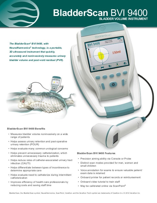
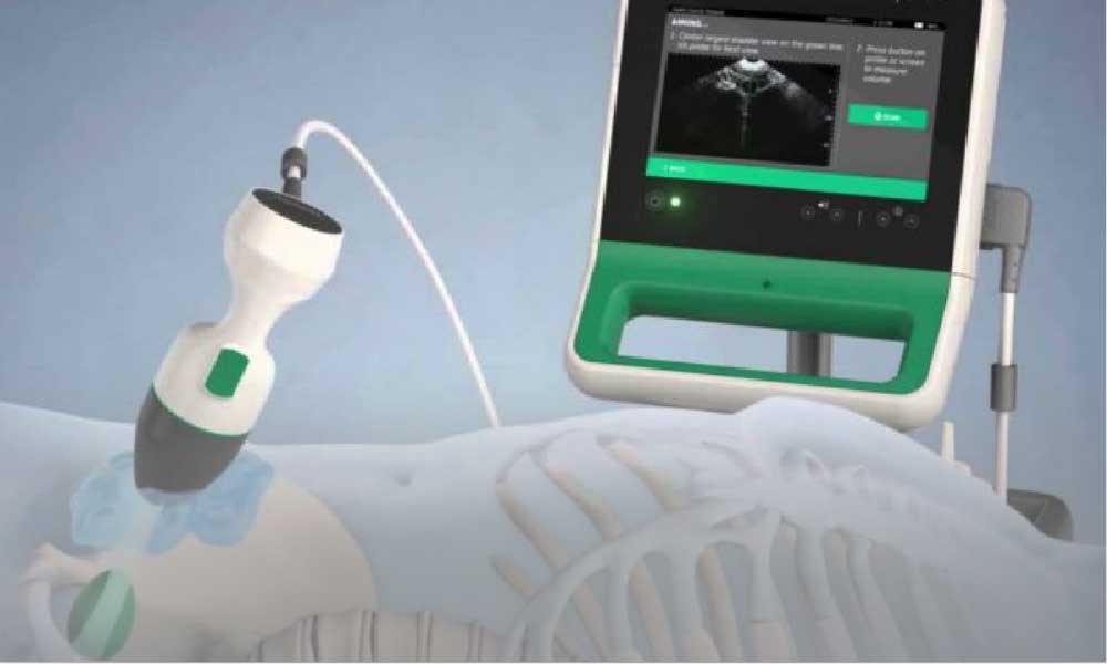
The bladder scanner is a portable, hand-held ultrasound device that can perform a quick, easy and non-invasive bladder scan. The scanner is equipped with an ultrasound probe and transducer that reflects the sound waves from the patients bladder into the scanner. The data is then transferred to a computer in a handheld unit for automatically calculating bladder volume. This ultrasonic procedure is also called Bladder Scanning or BladderScan, which is the brand name of the popular portable bladder scanner.
Bladder scanning is painless for the patient and eliminates the risks associated with unnecessary catheterization. The complete scanning lasts only a few minutes, does not require the intervention of an ultrasound technician and can prevent unnecessary invasive procedures.
Why Are Bladder Ultrasounds Done
Doctors order bladder ultrasounds when there’s a concern about bladder problems, such as trouble with peeing or daytime wetting.
A bladder ultrasound can show how much urine the bladder holds when it’s full and whether someone completely empties the bladder when peeing. It can show if theres anything unusual about the bladder, such as its size, the thickness of the bladder walls, and if there are blockages or kidney stones. A bladder ultrasound is often done along with a kidney ultrasound.
The doctor may ask that your child drink lots of fluids before the exam so that their bladder is full. The first image is done while your childs bladder is full. Then, your child will be asked to pee before more images are recorded.
Read Also: How Long Can You Live With Aggressive Bladder Cancer
How To Use A Bladder Scanner
wikiHow is a wiki, similar to Wikipedia, which means that many of our articles are co-written by multiple authors. To create this article, volunteer authors worked to edit and improve it over time. This article has been viewed 98,353 times.
A Pad Scan bladder scanner features new 3D sector probe and real-time ultrasound imaging algorithms that measures the urinary bladder volume and post-void residual quickly, safely, automatically and non-invasive. It is your ideal assistant to meet today Point-Of-Care challenges.
About The Ontario Health Technology Assessment Series

To conduct its comprehensive analyses, the Medical Advisory Secretariat systematically reviews available scientific literature, collaborates with partners across relevant government branches, and consults with clinical and other external experts and manufacturers, and solicits any necessary advice to gather information. The Medical Advisory Secretariat makes every effort to ensure that all relevant research, nationally and internationally, is included in the systematic literature reviews conducted.
The information gathered is the foundation of the evidence to determine if a technology is effective and safe for use in a particular clinical population or setting. Information is collected to understand how a new technology fits within current practice and treatment alternatives. Details of the technologys diffusion into current practice and information from practicing medical experts and industry, adds important information to the review of the provision and delivery of the health technology in Ontario. Information concerning the health benefits economic and human resources and ethical, regulatory, social and legal issues relating to the technology assist policy makers to make timely and relevant decisions to maximize patient outcomes.
Recommended Reading: Foods That Help Overactive Bladder
Benefits And Adverse Events
Ultrasound measurement of urine volumes compares favourably to manual palpation, which is imprecise, and to catheterization, which is the gold standard but is invasive and risk-laden. Ultrasound technology safely avoids trauma to the urinary tract and infection risks to the patient and attendant associated with catheterization. Using a portable bladder ultrasound device also eliminates the need to calculate bladder volumes manually from formulae, which reduces error introduction risks, and may avoid unnecessary catheterizations. Additionally, noninvasive biofeedback, toileting trials, and self-continence programs may be implemented based on portable bladder ultrasound measurements of urine volume and PVR. Lastly, as portable bladder ultrasound is a noninvasive transportable device, patient transfer is not necessary, and patient comfort and dignity are improved. Savings realized in nurses time, catheterization equipment, laundering, and medical imaging serve to offset the costs associated with implementing portable bladder ultrasound.
What Is It And When Should It Be Used
So what is it exactly and what is it used for? It is an ultrasound machine that uses sound waves to scan a patient’s bladder to determine how much urine they have inside. This is typically done if there is suspicion that they are retaining urine which can cause serious issues such as a urinary tract infection or even bladder rupture if not treated.
Note that just because they are urinating, that doesn’t necessarily mean they aren’t retaining. Retaining simply means they aren’t completely emptying out their bladder. To see if this is the case, the patient is scanned immediately after they urinate. This is called checking their post void residual. This is one reason why it is important to keep up with their intake and output .
After a patient has had their foley catheter removed, they need to urinate within a certain amount of time. At the hospital where I am employed, that time frame is 8 hours. If they still haven’t gone by then, we encourage them to go and if they are unable to, we bladder scan them. If they are retaining, there is usually an order to reinsert of the catheter or performing a straight cath.
How much urine in the bladder is considered too much? We typically have separate parameters ordered for each individual. They usually range between 250 and 350 mL , but it really depends on the patient. Of course generally speaking, the larger the patient, the larger the bladder.
Recommended Reading: Can You Treat A Bladder Infection Without Antibiotics
Why Perform A Bladder Scan
The aim of the post void bladder scan is to identify if the individual is emptying their bladder effectively when voiding, or if the volume of any residual urine left is significant.
Although urinary catheterisation has long been regarded as the gold standard in determining an accurate post void residual urine volume, it is invasive, sometimes uncomfortable, and carries a risk of infection or urethral trauma. It may still be used to determine PVR if a bladder scanner is not available or a client is not suitable for bladder scanning.
Bladder scanning has advantages over many other interventions, such as intermittent catheterisation, for measurement of post void residual volume, as it is simple and causes no discomfort to the patient/client.
In order to perform bladder scanning, health professionals must undergo training and be deemed competent to perform a bladder scan.
When a bladder scanner is available, estimation of residual urine or confirmation of effective bladder emptying should be a standard component of a continence assessment.
Note: Frailty, age or dementia are not reasons to not perform a bladder scan.
Clinical Practice Point. Remember that some individuals can present with a normal voiding pattern but have a significant post void residual.
What Is A Bladder Scanner
A bladder scanner is a hand-held, portable instrument that uses ultrasound imaging to perform a simple, non-invasive scan of the bladder. The computer in the device then receives the data and calculates urine volume. This gives the health practitioner an accurate idea of any necessary further steps.
Don’t Miss: Essential Oils For Bladder And Kidney Health
Action On Residual Urine
If residual urine is found an appropriate referral, usually to a consultant urologist, must be considered. But finding residual urine should not be the only factor to influence interventions such as a decision to catheterise. Renal function and lower urinary tract symptoms must be assessed and compared with the results of the scan before any intervention is contemplated .
According to the International Continence Society, 100ml or above is residual urine but a residue of 100ml is not likely to be acted on. Volumes of 300ml or more may require intervention, but varying amounts of residual urine may not cause any problems in relation to LUTS and renal function.
What Happens During My Exam
- You may be asked to change into a gown.
- A warm, non-scented, hypo-allergenic ultrasound gel will be applied to the area being scanned.
- Your technologist will ask you to lower or arrange your clothing so that the area of concern is exposed to apply gel and scan the area.
- The sonographer will move the transducer around the area between your hipbones and below your belly button to take images of your kidneys and bladder.
- The sonographer will ask you to perform various breathing techniques that aid in obtaining the best images of your organs.
- You may be asked to lie on your side, sit, or stand to bring your organs into a better position.
- You may experience mild to moderate pressure while the sonographer takes the images.
- You will be asked to empty your bladder at the end of the exam and then the sonographer will take more pictures of your bladder to measure the volume of remaining urine.
- The radiologist will review the images.
- The radiologist may come into the scan room after your exam to speak to you about your results. You will then be free to leave.
Don’t Miss: Overactive Bladder And Back Pain
How Much Does A Bladder Ultrasound Cost
If you have medical insurance, your copayment for a bladder ultrasound can vary or may even be free. Without insurance, the average cost of an ultrasound in the United States is between about $250 and $400.
If you have Medicare, an ultrasound may be covered under your Part A coverage if you have the procedure during an inpatient hospital stay.
At an outpatient facility, an ultrasound is covered by Medicare Part B. Your share of the cost can be between about $17 and $30 depending on where the test is done.
What Do I Need To Do To Prepare For My Bladder Ultrasound

Bladder ultrasound preparation is extremely minimal. Your ultrasound technician will let you know whether you need a full or empty bladder during your test. If you need to have a full bladder your doctor may ask you to drink many glasses of water before the test. If your bladder needs to be empty your ultrasound technician will simply ask you to use the restroom before your exam. If you have trouble keeping your bladder full, you may be asked to empty your bladder about an hour before your exam and then drink water once you get to the radiologists office so that the test can be done immediately after your bladder is filled. Therefore allowing you to not have to hold your urine as long.
You May Like: Can A Man Give A Woman A Bladder Infection
What Does A Bladder Ultrasound Show
A bladder ultrasound shows live pictures of your bladder, including your bladder wall and bladder pouches, also known as the diverticula. With such pictures, bladder ultrasounds can give doctors valuable information on variables like:
- Abnormalities with bladder size or thickness of bladder walls
- Bladder stones
- Cysts and tumors in the bladder
- Fluid collection
- How much urine the bladder can hold
- Urinary tract infections
Bladder ultrasounds use high-frequency sound waves delivered through a small, wand-shaped probe called a transducer. The transducer transmits the sound waves through the body and records the waves that echo back. The sound waves echo back once they hit a boundary between different tissues.
Such boundaries include those between fluid and soft tissue or soft tissue and bone. After recording the waves, the transducer sends the data to a nearby processing unit and monitor to display live pictures.
Magnetic Resonance Imaging Scan
Like CT scans, MRI scans show detailed images of soft tissues in the body. But MRI scans use radio waves and strong magnets instead of x-rays.
MRI images are very useful in showing cancer that has spread outside of the bladder into nearby tissues or lymph nodes. A special MRI of the kidneys, ureters, and bladder, known as an MRI urogram, can be used instead of an IVP to look at the upper part of the urinary system.
Recommended Reading: What To Drink For Overactive Bladder
Tests For Bladder Cancer
Bladder cancer is often found because of signs or symptoms a person is having. Or it might be found because of lab tests a person gets for another reason. If bladder cancer is suspected, exams and tests will be needed to confirm the diagnosis. If cancer is found, more tests will be done to help find out the extent of the cancer.
Why Is A Bladder Ultrasound Done
About a quarter of all people in the United States experience some level of incontinence, or the inability to hold urine in the bladder until you purposely release it.
There are many causes of incontinence, and it can be difficult for a doctor to pinpoint a reason for the problem just by asking you questions or examining the outside of your body.
The following symptoms may lead a doctor to order a bladder ultrasound:
- difficulty urinating
Don’t Miss: Where Is The Bladder Located
What Is A Bladder Ultrasound And How Is It Performed
A bladder ultrasound is a simple procedure that takes an image of the inside of the bladder, as well as the contents of it by using high-frequency sound waves transmitted through a transducer. This procedure is performed the same way in which many other ultrasounds are. A bladder ultrasound is a non-invasive procedure and not painful. It is performed by a sonographer who will put a clear gel on the patients lower abdomen between the belly button and pelvis, then the ultrasound technician will firmly press a transducer that will emit high-frequency sound waves. The way the sound waves bounce off of the patients body will create readable images that will allow doctors to detect information about the condition of the patients bladder.
Our Purpose Is To Transform Access To Education
We offer a diverse selection of courses from leading universities and cultural institutions from around the world. These are delivered one step at a time, and are accessible on mobile, tablet and desktop, so you can fit learning around your life.
We believe learning should be an enjoyable, social experience, so our courses offer the opportunity to discuss what youre learning with others as you go, helping you make fresh discoveries and form new ideas.You can unlock new opportunities with unlimited access to hundreds of online short courses for a year by subscribing to our Unlimited package. Build your knowledge with top universities and organisations.
Don’t Miss: Painful Bladder Syndrome Bladder Training
How To Do A Bladder Scan
There are several steps in a bladder scan procedure. To do a bladder scan, each of the steps must e followed carefully. Your doctor or medical practitioner will guide you through the steps of a bladder scan procedure. They will also inform you about any preparations that you may need to do. The steps for medical practitioners to follow in doing a bladder scan are
What Do The Results Mean

Simple types of bladder ultrasounds, called bladder scans, can deliver immediate results. These scans are usually used only to measure the amount of urine in your bladder. A diagnostic bladder ultrasound produces more complicated images about the size, fullness, and lining of the bladder.
A doctor might understand what the ultrasound is showing, but a radiologist will typically interpret the images and write a report for your doctor to review.
The doctor will make an official diagnosis after an ultrasound based on the report from the radiologist. Apart from overactive bladder, a bladder ultrasound may also be able to help diagnose bladder cancer.
After a diagnosis, the doctor can begin treatments or therapies to help your symptoms, such as medications or pelvic floor exercises. Sometimes, more testing may be needed.
If the doctor isnt certain of your diagnosis after a bladder ultrasound, they might order other tests.
Some other tests that can be used to examine the bladder include:
- urine lab testing
Recommended Reading: Can Overactive Bladder Cause Bloating
Why You Shouldnt Empty Your Bladder When Preparing For An Ultrasound
There are various reasons why an ultrasound may require a full bladder. In some cases, the ultrasound technologist may need you to have a full bladder so they can measure the bladder volume. Other times, the bladder fluid may provide enhanced visualization of surrounding pelvic organs.
Fluid serves as a visualization aid in ultrasound imaging because sound waves are denser and travel faster in water than in air. You experience these denser, faster sound waves whenever you dunk your head underwater and hear loud, muffled noises. The denser and faster sound waves enable greater detail with the pictures that ultrasound scans produce.
Its important to maintain a full bladder for ultrasounds with that requirement, as the accuracy of your results could be negatively impacted otherwise.